Radiologia Brasileira - Publicação Científica Oficial do Colégio Brasileiro de Radiologia
AMB - Associação Médica Brasileira CNA - Comissão Nacional de Acreditação
 Vol. 40 nº 5 - Sep. / Oct. of 2007
|
ORIGINAL ARTICLE
|
|
Acute pyelonephritis: frequency of findings in patients submitted to computed tomography |
|
|
Autho(rs): Flávio do Amaral Campos, George de Queiroz Rosas, Décio Goldenberg, Gilberto Szarf, Giuseppe D'Ippolito |
|
|
Keywords: Acute pyelonephritis, Kidney, Computed tomography, Reproducibility |
|
|
Abstract:
INTRODUCTION Acute pyelonephritis is a term currently recommended by the International Society of Uroradiology to define a renal inflammatory/infectious process(1). It is a common disease affecting renal parenchyma, interstice and pelvis(2), and most frequently occurs in the female adult population in the age range between 15 and 40 years(3). Most of times, the diagnosis is reached through clinical and laboratory tests(4). Imaging studies may be warranted if the disease presentation is confusing or atypical; in these cases, contrast-enhanced CT is recommended because of its high sensitivity and specificity(1,5,6). Its utilization is basically restricted to cases of suspicion of complications, for management of patients with an unfavorable clinical progression(5,7), or in the pursuance of some functional or morphological alteration of the urinary tract(3). Renal and perirenal tomographic signs found in cases of acute pyelonephritis have been already described in the literature(5,8-10). Few case reports have illustrated the finding of associated extrarenal alterations such as pleural effusion(11,12), gallbladder wall thickening, perivesicular fluid(13) and periportal edema(14,15). Until November/2006 there was no study in the literature describing a larger series of patients presenting with such extrarenal findings(7). Despite the fact that tomographic findings in patients with acute pyelonephritis are well known, the frequency of these findings, as well as CT reproducibility, still remains to be clearly established. This study was aimed at estimating the frequency of renal and extrarenal tomographic findings in patients with clinical laboratory signs of acute pyelonephritis, and evaluating the method reproducibility.
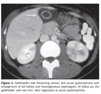
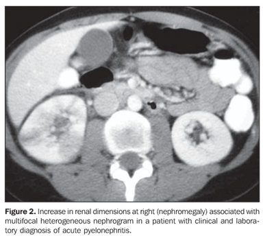
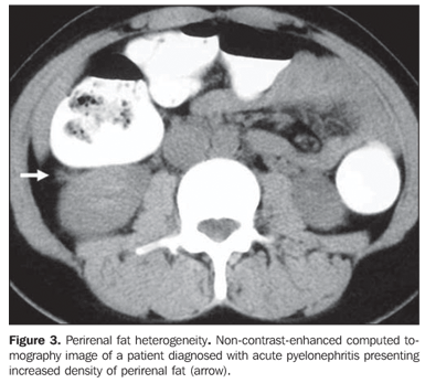
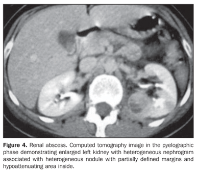
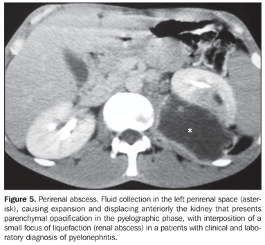
MATERIALS AND METHODS Abdominal CT images were selected in the period between February/2003 and February/2006 and included in the present retrospective, transversal and observational study. The patients evaluated had a clinical and laboratory diagnosis of acute pyelonephritis, and an initial tomographic report describing renal and extrarenal alterations compatible with AP. All of the images were acquired on a helical CT apparatus (Philips, Helicat Flash model) with thin, contiguous and interpolated slices, 5.5 mm-6.5 mm thickness (reconstruction with 5.0 mm) and 1-1.5 pitch, according to previously published technical parameters(8). The contrast-enhanced images were acquired after intravenous injection of iodinated, nonionic (Opriray, 320-320 mg/ml) contrast agent, at a dose of 2 ml/kg, up to a maximum volume of 200 ml, with an injection pump (rate 3 ml/s). All of the studies included a non-contrast enhanced phases, with nephrographic (75 seconds after contrast agent injection, and pyelographic (five minutes after contrast injection) images from the diaphragm up to the pubic symphysis. Of 56 CT studies initially selected, nine were excluded for being considered as inadequate due the absence of one of the main examination phases (non-contrast enhanced, nephrographic or pyelographic phases), surgery involving the urinary apparatus or findings compatible with chronic renal failure. A total of 47 patients - 43 women in the age range between 19 and 82 years (mean 31.3 years) and four men in the age range between 30 and 86 years (mean 54.3 years) - were studied. The images were blindly reviewed in a workstation by two radiologists, one of them with five-year experience in abdominal radiology (G.Q.R.), and the other in the 3rd year of residency in imaging diagnosis (F.A.C.), who have identified renal, perirenal and extrarenal alterations compatible with acute pyelonephritis. Cases of disagreement were reviewed by a specialist with 20-year experience (G.D.), aiming at establishing the absolute frequency of the classification for both acute pyelonephritis and tomographic signs. Acute pyelonephritis was classified into: a) unilateral or bilateral; b) focal or diffuse; c) with or without nephromegaly; d) complicated or non-complicate (with or without renal/perirenal abscess)(1). Renal, perirenal and extrarenal tomographic findings usually associated with acute pyelonephritis were analyzed, in an attempt to determine which of the kidneys was affected by the inflammatory process. The following renal and perirenal findings were taken into consideration: a) Nephromegaly - longitudinal renal axis > 11 cm(16) and/or focal or diffuse asymmetry of renal dimensions(5), considering that renal infection results in volume increase(17) (Figure 1); b) heterogeneous nephrogram - areas of hypoperfusion on the renal parenchyma(3) (Figure 2); c) perirenal fat heterogeneity - characterized by striated nephrogram, increased density of the perirenal fat and thickening of Gerota's fascia(5,8) (Figure 3); d) delay in contrast medium excretion - evaluation performed in the pyelographic phase where a delay in the intravenous iodinated contrast medium excretion is observed in the affected kidney(3); e) collecting system dilatation - asymmetrical dimensions of the pyelocaliceal system with enlargement of the affected kidney; f) nephrolithiasis and ureterolithiasis - presence of calculus in the collecting system(18); g) renal and perirenal abscess - area of liquefaction with well defined walls or pseudocapsula, possibly associated with gas attenuation or a thick aspect and density > 15 UH(5,8) (Figures 4 and 5).
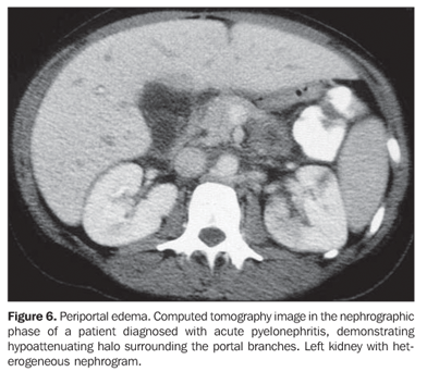
Findings of extra-renal involvement were: a) Gall bladder wall thickening or perivesicular fluid 3 4 mm-thick or presence of fluid adjacent to the gall bladder(1,9) (Figure 1); b) periportal edema - hypoattenuating halo involving the portal vein or its branches in contrast-enhanced phases(14,15) (Figure 6); c) Pleural effusion - any amount of fluid within the pleural space(11).
Statistical analysis of findings was performed, and interobserver agreement was calculated by means of kappa test (k)(20), considering the following levels: Insignificant agreement (k = 0.0-0.20), median (k = 0.21-0.40), moderate (k = 0.41-0.60), substantial (k = 0,61-0,80) and almost perfect (k = 0.81-1.00). Also, the chi-square test was utilized to identify the possible correlation between the presence of renal and perirenal findings with extra-renal findings. The significance level for null hypothesis rejection was established as < 0.05 (5%).
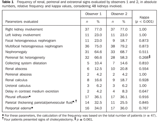
RESULTS The results analysis demonstrated findings of unilateral acute pyelonephritis in 46 cases (36 at right, and 10 at left), and bilateral em only one case, in a total of 48 kidneys affected by the disease. Acute pyelonephritis was classified as focal and multifocal, respectively, in 11/48 (23.0%) and 37/48 (77.0%) of cases; with and without nephromegaly, respectively, in 31/48 (64.6%) and 16/48 (33.4%) of cases; and complicated and non-complicated, respectively, in 10/48 (20.8%) and 38/48 (79.2%) of cases. The frequency of renal, perirenal and extrarenal findings of acute pyelonephritis for both observers 1 and 2 with respective kappa scores are shown on Table 1.
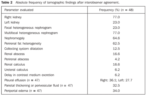
Amongst the studied findings, the only one presenting a median (k = 0.268) and statistically non-significant (p = 0.061) interobserver agreement was perirenal fat heterogeneity. In the chi-square test, a statistically significant correlation among renal, perirenal and extra renal findings could not be identified. Additionally, no statistically significant correlation could be established between the presence, separately or in conjunction, of one of the three extrarenal findings and the other findings of acute pyelonephritis. After analysis of the disagreeing cases, the respective frequencies of findings were defined as shown on Table 2.
DISCUSSION Acute pyelonephritis is a common disease. It is estimated that more than 250,000 new cases occur in United States each year(21). Its diagnosis is reached by means of clinical and laboratory tests; imaging studies such as ultrasound and computed tomography are utilized in case of diagnostic uncertainty or necessity to define associated complications(5). Main renal and perirenal tomographic signs found in cases of acute pyelonephritis are comprehensively described in the literature(5,8-10), but there is no reference to their frequency. The present study has tried to establish the distribution of acute pyelonephritis according to the previously proposed classification, the frequency of each tomographic finding, and the CT reproducibility, considering that the evaluation of the main findings is subjective. It is important to note that the total number of kidneys affected (n = 48) was utilized for calculating the frequency of renal and perirenal findings, and the number o patients (n = 47) was utilized for calculating the frequency of extrarenal findings. The present study has demonstrated a higher frequency of unilateral (46/47), multifocal (77.0%) acute pyelonephritis affecting the right kidney (77.0% of cases). Pleural effusion also was more prevalent in the right kidney (36.1%). No data was found in the literature to explain this fact. Heterogeneous nephrogram (either focal or multifocal) was observed in all of the cases studied, and was considered as the main tomographic finding indicative of acute pyelonephritis, and also the most objective because of the excellent interobserver agreement (k = 0.873). However, it is known that heterogeneous nephrogram is not pathognomonic of acute pyelonephritis, and may occur in cases where a low-osmolarity contrast agent is utilized in dehydrated patients or in other clinical circumstances like in patients with renal infarction or acute tubular necrosis(6). Nephromegaly was present in two-thirds of cases. It is important to note that an objective data (the longest renal axis) and a subjective data (renal asymmetry) were considered in conjunction. If only the maximum renal diameter was considered as indicative of nephromegaly, this finding would have been found in only one-third (31%) of patients. Acute pyelonephritis was classified as complicated, i.e., associated with renal and/or perirenal abscess in about 21% of cases. The significance of the renal abscess detection is controversial. In the literature there are studies demonstrating that small renal abscesses may resolve only with a clinical treatment, with no need of an invasive procedure(1). However, the treatment strategy only should be defined after the characterization of the abscess. Interobserver disagreement was observed in four cases of suspicion of renal abscess. All of these cases were reviewed as heterogeneous nephrogram with interposition of small hypoattenuating foci, which has generated a doubtful interpretation, although with no impact on the management of the patients, since the lesions were small undrainable abscesses. Ureteral obstruction leading to collecting system stasis may be a triggering factor for acute pyelonephritis(5). In the present casuistic, ureteral calculi were detected only in three cases (6.2%), and upstream collecting system dilatation in only one. Also in this case, association between the presence of renal calculi and renal abscesses was not observed. Perirenal fat heterogeneity was the sole finding among the other studied that did not achieved a significant interobserver agreement. This finding was considered as the most subjective among the others, so its reproducibility was low. One of the reasons for this disagreement may be related to the "window" utilized in the examination. It is important to note that this is a non-specific finding also observed in cases of previous inflammatory process sequela in vascular disease, trauma(6) or urinary tract obstruction(22). All of the other (renal, perirenal and extrarenal) findings showed a statistically significant interobserver agreement (p = 0.005) ranging between moderate and almost perfect, despite the different levels of experience of the observers and the subjectivity in the evaluation of some findings. The presence of extrarenal alterations, particularly those affecting the liver and the biliary tract should be highlighted. Until recently, there was only sporadic reports on cases of patients with acute pyelonephritis and gallbladder wall thickening or perivascular fluid(13) and periportal edema(14,15). Periportal edema had already been described for other conditions such as cirrhosis, hepatitis, liver tumors, biliary tract obstruction, liver and bone marrow transplant, heart failure, abdominal trauma in the pediatric population(14,23-25), and even in acute pyelonephritis(7,15). In the present study this was a relatively frequent finding, present in one-third of cases. There are several causes for gallbladder wall thickening/perivesicular fluid, except alterations of the gallbladder itself, such as ascites, hypoalbuminemia, perivesicular abscess, peptic ulcer, pancreatitis, hepatitis, cirrhosis, portal hypertension, chronic heart and renal failure(13). Acute pyelonephritis also has been described as a cause for gallbladder wall thickening(7,13). The present study identified this finding or perivesicular fluid in one-third of cases. It is important to note that both the acute cholecystitis and acute may cause of pain in the right hypochondrium associated with gallbladder parietal thickening or perivesicular fluid, and should be taken into consideration in the differential diagnosis. There is a belief that extrarenal alteration occur because of multiple factors related to the septic condition and metabolic disorders present in the setting of infectious processes(7). In the largest casuistic so far (21 patients) describing extrarenal alterations in patients with acute pyelonephritis(7), the authors have found a frequency different from the one found in the present study, maybe because of the inclusion and selection criteria adopted. Additional studies will be necessary for a more accurate estimation of the true frequency of these findings. It is important to note that the majority of the patients in the present study (89%) presented alterations directly related to the acute pyelonephritis presentation, allowing to consider the positivity of this tomographic findings as a result of the renal inflammatory and infectious condition, as already identified by other authors(7). However, it is important to note that some of these findings, perirenal heterogeneity and collecting system dilatation observed in patients with excretory system obstruction, for example, are non-specific of acute pyelonephritis(26). On the other hand, it is also important to note that, for the first time, the reproducibility of these tomographic findings in patients with acute pyelonephritis was established, giving the method higher reliability, independently from the investigator experience. One of the shortcomings of the present study is the fact that it has not included all the patients assisted with suspicion for acute pyelonephritis, and therefore the absolute frequency of tomographic alterations could not be determined in this group of patients. The main hindrance in this type of study would be to submit a patient to an unnecessary examination, considering the clinical presentation of acute pyelonephritis which frequently does not require supplementary imaging studies. Additionally, tomographic findings were not correlated with clinical presentation, laboratory tests results and clinical progression of the disease, which could define prognostic signs similarly to those described in other clinical situations. A new prospective study is going to be developed by the authors to establish the correlation between tomographic findings and clinical progression. The present study demonstrated that, in patients submitted to CT scan, the frequency of the different tomographic findings suggestive of acute pyelonephritis is high, and heterogeneous nephrogram is the most frequent and reliable. Perirenal and extrarenal alterations were observed in up to two-thirds of cases included in the present study. Despite their subjectivity, tomographic signs of acute pyelonephritis present high reproducibility, ratifying the CT value in the evaluation of this group of patients.
REFERENCES 1.Talner LB, Davidson AJ, Lebowitz RL, Dalla Palma L, Goldman SM. Acute pyelonephritis: can we agree on terminology? Radiology 1994;192: 297–305. [ ] 2.Cotran SC, Kumar V, Collins T. Rim. In: Cotran SC, Kumar V, Collins T, editores. Robbins Patologia estrutural e funcional. 6ª ed. Rio de Janeiro, RJ: Guanabara Koogan, 2000;874–875. [ ] 3.Gold RP, McClennan BL. Acute infections of renal parenchyma. In: Pollack HM, editor. Clinical urography. 2nd ed. Philadelphia, PA: Saunders, 1990;799–821. [ ] 4.Papanicolaou N, Pfister RC. Acute renal infections. Radiol Clin North Am 1996;34:965–995. [ ] 5.D'Ippolito G, Abreu Jr L, Borri ML, Galvão Filho MM, Hartmann LGC, Wolosker AMB. Pielonefrite aguda: classificação, nomenclatura e diagnóstico por imagem. Rev Imagem 2005;27: 183–194. [ ] 6.Stunell H, Buckley O, Feeney J, Geoghegan T, Browne RF, Torreggiani WC. Imaging of acute pyelonephritis in the adult. Eur Radiol 2007;17: 1820–1828. Epub 2006 Aug 26. [ ] 7.Zissin R, Osadchy A, Gayer G, Kitay-Cohen Y. Extrarenal manifestations of severe acute pyelonephritis: CT findings in 21 cases. Emerg Radiol 2006;13:73–77 [ ] 8.Kawashima A, Sandler CM, Goldman SM, Raval BK, Fishman EK. CT of renal inflammatory disease. RadioGraphics 1997;17:851–866. [ ] 9.Kawashima A, LeRoy AJ. Radiologic evaluation of patients with renal infections. Infect Dis Clin North Am 2003;17:433–456. [ ] 10.Rabushka LS, Fishman EK, Goldman SM. Pictorial review: computed tomography of renal inflammatory disease. Urology 1994;44:473–480. [ ] 11.Berkman N, Liss H, Kramer MR. Pyelonephritis as a cause of pleural effusion. Respiration 1996; 63:384–386. [ ] 12.Wang IK, Chuang FR, Chang HY, Lin CL, Yang CT. Acute pyelonephritis associated with transudative pleural effusion in a middle-aged woman without urinary tract obstruction. Med Princ Pract 2006;15:309–311. [ ] 13.Talarico HP, Rubens D. Gallbladder wall thickening in acute pyelonephritis. J Clin Ultrasound 1990;18:653–657 [ ] 14.Lawson TL, Thorsen MK, Erickson SJ, Perret RS, Quiroz FA, Foley WD. Periportal halo: a CT sign of liver disease. Abdom Imaging 1993;18:42–46. [ ] 15.Zissin R, Kots E, Rachmani R, Hadari R, Shapiro-Feinberg M. Hepatic periportal tracking associated with severe acute pyelonephritis. Abdom Imaging 2000;25:251–254. [ ] 16.Emamian SA, Nielsen MB, Pedersen JF, Ytte L. Kidney dimensions at sonography: correlation with age, sex, and habitus in 665 adult volunteers. AJR Am J Roentgenol 1993;160:83–86. [ ] 17.Gold RP, McClennan BL, Rottenberg RR. CT appearance of acute inflamatory disease of the renal interstitium. AJR Am J Roentgenol 1983; 141:343–349. [ ] 18.Lee Jr FT, Thornbury JR. O trato urinário. In: Juhl JH, Crummy AB, Kuhlman JE, editores. Interpretação radiológica. 3ª ed. Rio de Janeiro, RJ: Guanabara Koogan, 2000;543–624 [ ] 19.Laing FC. A vesícula biliar e os ductos biliares. In: Rumack CM, editor. Tratado de ultra-sonografia diagnóstica. 2ª ed. Rio de Janeiro, RJ: Guanabara Koogan, 1999;149–190. [ ] 20.Landis JR, Koch GG. The measurement of observer agreement for categorical data. Biometrics 1977;33:159–174. [ ] 21.Ramakrishnan K, Scheid DC. Diagnosis and management of acute pyelonephritis in adults. Am Fam Physician 2005;71:933–942. [ ] 22.Rucker CM, Menias CO, Bhalla S. Mimics of renal colic: alternative diagnoses at unenhanced helical CT. RadioGraphics 2004;24 Suppl 1:S11–28. [ ] 23.Aspestrand F, Schrumpf E, Jacobsen M, Hanssen L, Endresen K. Increased lymphatic flow from the liver in different intra- and extrahepatic diseases demonstrated by CT. J Comput Assist Tomogr 1991;15:550–554. [ ] 24.Koslin DB, Stanley RJ, Berland LL, Shin MS, Dalton SC. Hepatic perivascular lymphedema: CT appearance. AJR Am J Roentgenol 1988;150: 111–113. [ ] 25.Patrick LE, Ball TI, Atkinson GO, Winn KJ. Pediatric blunt abdominal trauma: periportal tracking at CT. Radiology 1992;183:689–691. [ ] 26.Galvão Filho MM, D'Ippolito G, Hartmann LGC, et al. O valor da tomografia computadorizada helicoidal sem contraste na avaliação de pacientes com dor no flanco. Radiol Bras 2001;34:129–134. [ ]
Received December 15, 2006. Accepted after revision February 27, 2007.
* Study developed in the Department of Imaging Diagnosis at Universidade Federal de São Paulo/Escola Paulista de Medicina (Unifesp/EPM), and Hospital São Luiz, São Paulo, SP, Brazil. |
|

Av. Paulista, 37 - 7° andar - Conj. 71 - CEP 01311-902 - São Paulo - SP - Brazil - Phone: (11) 3372-4544 - Fax: (11) 3372-4554