Radiologia Brasileira - Publicação Científica Oficial do Colégio Brasileiro de Radiologia
AMB - Associação Médica Brasileira CNA - Comissão Nacional de Acreditação
 Vol. 41 nº 6 - Nov. / Dec. of 2008
|
ORIGINAL ARTICLE
|
|
Differentiation between simple cyst and hepatic hemangioma utilizing T2-weighted magnetic resonance imaging with gradient-echo (b-FFE) technique |
|
|
Autho(rs): Carolina Valente Burim, Luiz Pecci Neto, Fabiola Goda Torlai, Dario Ariel Tiferes, Giuseppe D'Ippolito |
|
|
Keywords: Liver, Cyst, Hemangioma, Magnetic resonance imaging |
|
|
Abstract:
IBiomedician, Fellow PhD degree, Departamento de Diagnóstico por Imagem da Universidade Federal de São Paulo/Escola Paulista de Medicina (Unifesp/EPM), São Paulo, SP, Brazil
INTRODUCTION Magnetic resonance imaging (MRI) has been frequently utilized for detecting and characterizing hepatic nodules, among other applications in the evaluation of abdominal diseases(1). Hepatic nodules are frequently found on abdominal MRI studies and must be characterized. In some cases MRI results can be correlated with those previously found at ultrasonography (US) and computed tomography (CT), allowing a complementary diagnosis. In other cases, these methods are not available or simply have not been performed, so MRI becomes the single source of information for guiding the hepatic nodule characterization. Amongst the hepatic nodules, hemangiomas and simple cysts are the most frequently found and can be present in up to 20% in cases of autopsy(2). At non-contrast-enhanced MRI, hepatic hemangiomas and simple cysts present similar behavior in signals, characterized by nodular, hypointense images on T1-weighted sequences and hyperintense on T2-weighted sequences, with no significant signal loss when long echo times (TE > 140 ms) are used(3), differently from other types of hepatic nodules such as hepatocarcinomas or metastases where signal loss can be observed as TE is extended on T2-weighted images(3). Because of the similarity between the signals of these lesions, intravenous paramagnetic contrast (gadolinium) injection is required for differentiation between cysts and hemangiomas(1,4). Despite the safety and low toxicity of the gadolinium-based paramagnetic contrast agents, especially as compared with iodinated contrast agents, the utilization of this type of contrast agent increases the cost of the study. Additionally, most recently, an association between the utilization of gadolinium and the development of systemic nephrogenic fibrosis has been reported in patients with chronic renal failure, thus limiting in some way the utilization of gadolinium-based contrast agents(5). Some authors have successfully devoted themselves to the study of the differentiation between hepatic cysts and hemangiomas based only on T2-weighted sequences, without the utilization of paramagnetic contrast injections(4,6). Recently a pulse gradient-echo sequence was developed with a balanced gradient structure (balanced-FFE – b-FFE), allowing the acquisition of T2-weighted sequences in short acquisition times and without the presence of motion artifacts resulting from respiration(7). The authors have observed that this sequence would allow the differentiation between hepatic hemangiomas and simple cysts without the necessity of intravenous contrast-enhancement, because of the hemangiomas signal loss, differently from the behavior presented by simple cysts. The present study was aimed at evaluating the role of MRI T2-weighted images in the differentiation between hepatic simple cysts and hemangiomas.
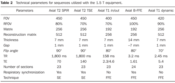
MATERIALS AND METHODS In the period between February 2005 and February 2006, the authors developed a prospective, observational, cross-sectional, double-blinded study analyzing gradient-echo b-FFE abdominal MRI images of 52 patients with a total of 91 lesions corresponding to 34 simple cysts and 57 hepatic hemangiomas diagnosed by contrast-enhanced MRI. The group of patients included 36 women and 16 men with ages ranging from 23 to 80 years (mean = 47.5 years). The research protocol of the present study was approved by the Committee for Ethics in Research (Project No. 0922/05) of Universidade Federal de São Paulo/Hospital São Paulo, and by the Committee for Ethics in Research (Project No. 092/2005) of Beneficência Médica Brasileira S/A – Hospital e Maternidade São Luiz. The studies were performed in a 1.0 T MRI equipment Gyroscan NT10 (Philips Medical Systems; Best, The Netherlands) (45 patients with 30 cysts and 48 hemangiomas) and a 1.5 T Intera ACS NT15 (Philips Medical Systems; Best, The Netherlands) (7 patients with 4 cysts and 9 hemangiomas), respectively with body and synergy coils. The paramagnetic contrast agent was administered through a Medrad injection pump to patients after four-hour fasting. All of them were intravenously administered butylscopolammonium bromide (1 ml diluted in 10 ml sodium solution) immediately before the examination through a Gelco 18-20 gauge catheter into an antecubital calibrous vein to allow a safe and adequate contrast injection. All of the patients received a dose of 0.2ml/kg of paramagnetic contrast agent (Gd-DTPA), at an injection rate of 2 ml/s. Contrast-enhanced images were acquired 20 seconds (arterial phase), 60 seconds (portal phase) and five minutes (equilibrium phase) after the intravenous contrast injection was initiated. The sequences utilized with the 1.0 T and 1.5 T equipments as well as the respective technical parameters are summarized on Tables 1 and 2.
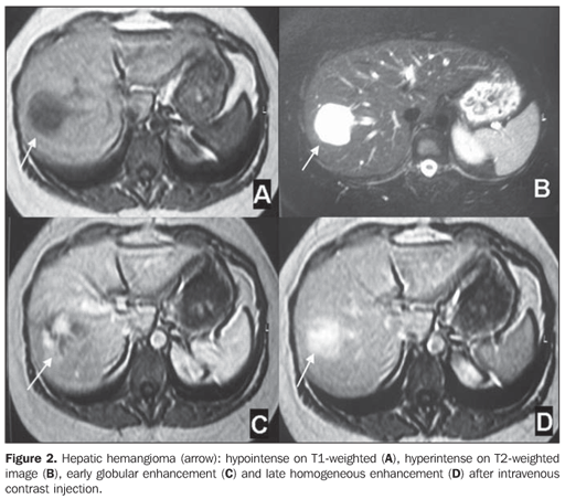
The golden-standard for determining the nature of the nodule (simple cyst or hemangioma) was defined by a radiologist with an extensive experience in abdominal MRI who analyzed all the T1- and T2-weighted with short and long TE, the images of the b-FFE sequences, and those acquired after paramagnetic contrast injection. Simple cysts were those lesions with the following MRI findings: well-defined hypointense nodule at T1-weighted, and hyperintense at T2-weighted images, with no apparent signal loss with the longer echo times (TE = 140 ms), homogeneous contents, absence of septa or vegetations, and with no contrast-enhancement after paramagnetic contrast injection(8) (Figure 1). Hemangiomas were those lesions with the following MRI findings: well-defined hypointense nodule at T1- and hyperintense at T2-weighted sequences, even with the longest echo times (TE = 140 ms) and peripheral globular enhancement with centripetal fill-in or early and progressive homogeneous enhancement, without distortion of adjacent vessels or hepatic capsule retraction(8) (Figure 2).
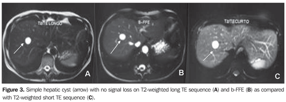
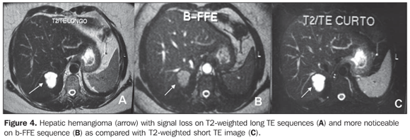
Images analysis The images were interpreted by two independent observers with one-year education in abdominal MRI, who had no previous knowledge of the US, CT and contrast-enhanced MRI results. The analysis of the images was performed on an EasyVision workstation (Philips Medical Systems; Best, The Netherlands) or on the MRI equipment console, specifically focusing on the localization and diameter of previously identified nodules. Only the T2-weighted sequences were evaluated by the two observers who subjectively classified the hepatic lesions into either simple cysts or hemangiomas according to the findings at T2weighted images, adopting the sequences obtained with short TE (TE = 70–90 ms), long TE (TE = 140 ms) and b-FFE as parameters. Simple cysts were those lesions that did not present signal loss on the long TE or b-FFE sequences as compared with the short TE sequences (Figure 3), and hemangiomas were those with apparent signal loss on these sequences (Figure 4).
The images were independent and blindly analyzed by the two observers at three different moments with a minimum 15-day interval, as follows: moment 1 (M1) — comparison between results with short-TE and long TE for determining the value of long TE sequences; moment 2 (M2) —first comparison between short TE and b-FFE sequences for determining the value of the b-FFE sequence and allowing the calculation of the interobserver agreement; moment 3 (M3) — second comparison between short TE and b-FFE sequences to allow the calculation of the interobserver agreement. Statistical analysis Intra- and interobserver agreements were calculated by means of the kappa index (κ) and the significance level (p) adopted was 0.05 (α = 5%)(15).
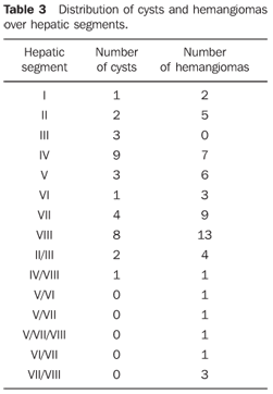
RESULTS Hepatic nodules localization and dimensions The cysts evaluated in the present study ranged from 0.5 to 6.5 cm in diameter (mean 1.89 cm). The distribution of the cysts and over hepatic segments is demonstrated on Table 3.
The hemangiomas diameter ranged between 0.8 and 11 cm (mean 2.62 cm). The distribution of the hemangiomas over hepatic segments is demonstrated on Table 3. Value of T2-weighted sequences The agreement between the analysis of TSE sequence with long TE (M1) and the golden-standard was poor and non-significant for both observers (κ: 0.10 and κ: 0.00; p = 0.196 and p= 0.883; non-significant). The agreement between the b-FFE (M2 and M3) sequence and the golden-standard ranged from substantial (κ: 0.62 and 0.71) to almost perfect (κ: 0.86) and was statistically significant (p < 0.001*). The interobserver agreement was considered as substantial in both moments of the analysis (κ: 0.62–0.70). The intra-observer agreement in relation to the evaluations at moments M2 and M3 was considered as almost perfect (κ: 0.85–0.91) for both observers. Statistical differences between the groups of patients evaluated with the 1.0 T and 1.5 T systems could not be calculated because of the small number (n = 7) of studies performed with the 1.5 T MRI system.
DISCUSSION MRI has been frequently utilized for detecting and characterizing hepatic nodules among other applications in the evaluation of abdominal diseases(1). In this context, hepatic nodules are most frequently found, the benign ones corresponding to simple cysts present in 2%–7% of the general population, and hemangiomas present in up to 20% of cases of autopsy(2,8). Generally these nodules are asymptomatic, being incidentally found on imaging studies. Diagnosis confirmation in these cases is most frequently achieved by means of a combination of imaging methods including US, CT and/or MRI(4,8). Hepatic simple cysts and hemangiomas are considered as benign lesions, representing a diagnostic challenge considering that, at imaging studies, these lesion could be mistaken with primary of secondary malignant neoplasms, leading to unnecessary surgical procedures or incorrect cancer staging(8). MRI is considered as a reliable method for detecting and characterizing hepatic lesions. Advantages of this method include multiplanar imaging capability, higher specificity due to T2-weighted images, and absence of ionizing radiation and more nephrotoxic iodine-based contrast agents like those utilized at CT(8). The strategy adopted in MRI studies for differentiation between benign and malignant hepatic nodules combines a visual evaluation of signal intensity on dynamic contrast-enhanced, heavily T2-weighted and T1-weighted images. Among benign nodules, cysts and hemangiomas are most frequently found, both presenting high signal intensity on T2-weighted sequences with longer echo times (ex.: TE = 140 ms), differently from metastases and hepatocarcinoma, which present with lower signal intensity on this type of sequence(1,3). On the other hand, the paramagnetic contrast injection allows the completion of this differentiation through the analysis of the hepatic nodule enhancement pattern(9). So, considering the similarity of signal intensity between cysts and hemangiomas, paramagnetic contrast (Gd-DTPA) injection becomes necessary for differentiation between these lesions. Cysts do not enhance after administration of Gd-DTPA, and hemangiomas present an early, discontinuous peripheral, progressive and centripetal enhancement(2,8). Despite of the safety and low toxicity, especially as compared with iodinated contrast agents, the utilization of a paramagnetic contrast agent results in an increase in the cost of the study. Additionally, recent studies have demonstrated an association between the utilization of gadolinium and the development of nephrogenic systemic fibrosis in patients with chronic renal failure(5). Some limitations related to the utilization of gadolinium-based contrast agents, and the possibility of MRI becoming a completely non-invasive method for differentiating hepatic cysts from hemangiomas have lead some authors to perform such differentiation without utilizing intravenous contrast injection, with quite significant rates of success(4,6). However, it is important to observe that these authors have not evaluated the accuracy of the method through the calculation of intra-observer agreement, raising the necessity of a further prospective evaluation of a higher number of lesions like the present study. Additionally, the sequences performed required several acquisitions and breath-holds for evaluating the whole liver, which could extend the acquisition time, besides failing in the detection of all the hepatic lesions present(6). A sequence available in the majority of modern MRI systems (b-FFE) was utilized in the present study, with short acquisition time (< 60 seconds), low sensitivity to artifacts and not requiring breath-hold or respiratory synchronization, a method feasible for patients with limited ventilator capacity(10). The present prospective study was designed to allow a comparison of the b-FFE sequence with the golden-standard in the characterization of hemangiomas, i.e., TSE with long TE(4,11,12). So, the authors sought to establish the role of this sequence in the differentiation between hemangiomas and cysts, which had not been proposed in the literature yet. For this purpose, MRI was established as a golden-standard encompassing all the sequences adopted in the routine investigation of hepatic nodules, including contrast-enhanced sequences during arterial, portal venous and equilibrium phases(9). This golden-standard has been utilized by the authors of the present study and by others, and also has been accepted in the literature as a reliable method for evaluating the effectiveness of new MRI sequences for detecting or characterizing hepatic nodules(6,12). The results of the present study demonstrated that the b-FFE sequence is not only more effective than TSE with long TE, but also is more accurate, with a considerably high inter-observer agreement. It is important to observe that, in the present sample, the authors evaluated nodules with up to 0.5 cm in diameter, localized in different segments of the liver, with no difficulty in differentiating cysts from hemangiomas, even in the case of nodules < 1.0 cm. In cases of small hepatic nodules (< 1.0 cm) MRI sequences traditionally utilized may present a low accuracy(13). In the present study, both the observers had only one year of experience after the period of residency in imaging diagnosis, which corroborates the simplicity of the method and the short learning curve. It took only one training session to instruct the two observers about the criteria adopted in the images analysis. Some criticisms could be raised against the present study. Firstly, only hemangiomas considered as typical, corresponding to about 80% of all hepatic hemangiomas, were evaluated(14). Results including atypical hemangiomas such as sclerosing hemangiomas or hemangiomas with intralesional hemorrhage, are still to be known. On the other hand, considering the utilization of MRI as a goldenstandard, atypical hemangiomas could not be included because its etiology could not be confirmed unless a long-term follow-up was undertaken or a biopsy was performed for anatomopathological analysis. It is important to note that hemangiomas can develop with time and evolutive controls not always are reliable to prove the etiology of the lesion(14). Among the patients of the present sample, those with a history of malignant neoplasm were excluded, considering that some tumor result in cystic hepatic metastasis that may manifest as atypical cysts, with intracystic hemorrhage or a thick content of other nature(15). Therefore, only patients with simple hepatic cysts were evaluated. Probably, the results would be different if patients with complex hepatic cysts had been included, considering that the behavior of these lesions on T2-weighted images is quite different from the simple cysts(15). Also, the authors could not determine the results for patients with hepatic steatosis, since these patients were not included in the present sample. This variable should still be evaluated, considering that hepatic steatosis may affect the hepatic parenchyma signal intensity on T2-weighted images with fat-suppression (SPIR), like those utilized in the present study(16). The evaluation of an objective parameter, for example, the nodule/liver signal ratio could add subsidiary results for the differentiation between cysts and hemangiomas on the different sequences utilized and this is a reason for continuing the present study whose results should be reported in a near future. Finally, the results of the present study indicate that the bFFE sequence plays a significant role in the routine MRI evaluation of the liver, considering that this is a fast sequence, with a relatively low sensitivity to motion or respiratory artifacts and high effectiveness and accuracy in the differentiation between simple cysts and hepatic hemangiomas. Despite not requiring the utilization of paramagnetic contrast agent for this differentiation, gadolinium-based contrast agent is still necessary in liver MRI for detection and characterization of a wide range of focal hepatic lesions(9,17,18).
REFERENCES 1. Cieszanowski A, Szeszkowski W, Golebiowski M, et al. Discrimination of benign from malignant hepatic lesions based on their T2-relaxation times calculated from moderately T2-weighted turbo SE sequence. Eur Radiol. 2002;12:2273–9. [ ] 2. Karhunen PJ. Benign hepatic tumours and tumour like conditions in men. J Clin Pathol. 1986;39:183–8. [ ] 3. D'Ippolito G, Tiferes DA. Estudo quantitativo da intensidade de sinal do hemangioma hepático: um novo parâmetro utilizado em ressonância magnética de alto campo. Radiol Bras. 1995;28:125–30. [ ] 4. Kiryu S, Okada Y, Ohtomo K. Differentiation between hemangiomas and cysts of the liver with single-shot fast-spin echo image using short and long TE. J Comput Assist Tomogr. 2002;26:687–90. [ ] 5. Broome DR, Girguis MS, Baron PW, et al. Gadodiamide-associated nephrogenic systemic fibrosis: why radiologists should be concerned. AJR Am J Roentgenol. 2007;188:586–92. [ ] 6. Sasaki K, Ito K, Koike S, et al. Differentiation between hepatic cyst and hemangioma: additive value of breath-hold, multisection fluid-attenuated inversion-recovery magnetic resonance imaging using half-Fourier acquisition single-shot turbo-spin-echo sequence. J Magn Reson Imaging. 2005;21:29–36. [ ] 7. Keogan MT, Edelman RR. Technologic advances in abdominal MR imaging. Radiology. 2001;220: 310–20. [ ] 8. Horton KM, Bluemke DA, Hruban RH, et al. CT and MR imaging of benign hepatic and biliary tumors. Radiographics. 1999;19:431–51. [ ] 9. Martin DR, Danrad R, Hussain SM. MR imaging of the liver. Radiol Clin North Am. 2005;43: 861–86, viii. [ ] 10. Meirelles GSP, Tiferes DA, D'Ippolito G. Pseudolesões hepáticas na ressonância magnética: ensaio iconográfico. Radiol Bras. 2003;36:305–9. [ ] 11. Li W, Nissenbaum MA, Stehling MK, et al. Differentiation between hemangiomas and cysts of the liver with nonenhanced MR imaging: efficacy of T2 values at 1.5 T. J Magn Reson Imaging. 1993;3:800–2. [ ] 12. Tiferes DA, D'Ippolito G, Szejnfeld J. Ressonância magnética dos hemangiomas hepáticos: avaliação das características morfológicas e quantitativas. Radiol Bras. 2003;36:1–9. [ ] 13. Kim KW, Kim AY, Kim TK, et al. Small (< or = 2 cm) hepatic lesions in colorectal cancer patients: detection and characterization on mangafodipir trisodium-enhanced MRI. AJR Am J Roentgenol. 2004;182:1233–40. [ ] 14. D'Ippolito G, Appezzato LF, Ribeiro ACR, et al. Apresentações incomuns do hemangioma hepático: ensaio iconográfico. Radiol Bras. 2006;39: 219–25. [ ] 15. Mortelé KJ, Ros PR. Cystic focal liver lesions in the adult: differential CT and MR imaging features. Radiographics. 2001;21:895–910. [ ] 16. Qayyum A, Goh JS, Kakar S, et al. Accuracy of liver fat quantification at MR imaging: comparison of out-of-phase gradient-echo and fat-saturated fast spin-echo techniques – initial experience. Radiology. 2005;237:507–11. [ ] 17. D'Ippolito G, Abreu Jr L, Borri ML, et al. Apresentações incomuns do hepatocarcinoma: ensaio iconográfico. Radiol Bras. 2006;39:137–43. [ ] 18. Machado MM, Rosa ACF, Lemes MS, et al. Hemangiomas hepáticos: aspectos ultra-sonográficos e clínicos. Radiol Bras. 2006;39:441–6. [ ] Received October 3, 2007. Accepted after revision April 23, 2008. * Study developed in the Departamento de Diagnóstico por Imagem da Universidade Federal de São Paulo/Escola Paulista de Medicina (Unifesp/EPM), and Serviço de Ressonância Magnética do Hospital São Luiz, São Paulo, SP, Brazil. |
|
Av. Paulista, 37 - 7° andar - Conj. 71 - CEP 01311-902 - São Paulo - SP - Brazil - Phone: (11) 3372-4544 - Fax: (11) 3372-4554

