Radiologia Brasileira - Publicação Científica Oficial do Colégio Brasileiro de Radiologia
AMB - Associação Médica Brasileira CNA - Comissão Nacional de Acreditação
 Vol. 41 nº 4 - July / Aug. of 2008
|
CASE REPORT
|
|
Diabetic mastopathy: an unusual differential diagnosis: a case report |
|
|
Autho(rs): Simone Elias, Marina Celli Francisco, Cláudio Kemp, Beatriz Daou Verenhitach, Fabiano Celli Francisco, Maria del Carmen M. Wolgien |
|
|
Keywords: Diabetic mastopathy, Diabetes mellitus, Breast benign disease |
|
|
Abstract:
IPhD, MD, Mastologist, Diagnostic Unit, Division of Mastology, Universidade Federal de São Paulo/Escola Paulista de Medicina (Unifesp/EPM), São Paulo, SP, Brazil
INTRODUCTION Breast is not classically included among organs affected by diabetic complications; however, a pathological manifestation defined as diabetic mastopathy may rarely occur (less than 1% of benign diseases)(1), affecting up to 13% of patients with diabetes mellitus type I, at some moment in the course of the disease. Diabetic mastopathy was firstly described in 1984 by Soler & Khardori, as a benign disease clinically simulating a breast carcinoma(2). Its pathogenesis is still to be completely known, but it is believed that this condition develops as an autoimmune reaction induced by the hyperglycemia, which would result in an increased production of collagen(1), with consequential lymphocytic infiltrate with B lymphocytes against the final products of the abnormal extracellular matrix glycosylation(3).
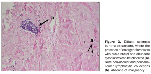
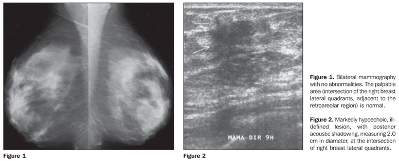
CASE REPORT A female 35-year-old patient complaining of a lump found during self-examination, at the intersection of the lateral quadrants of her right breast, near the retroareolar region. Insulin-dependent, diabetic (diabetes mellitus) patient since she was 16 years old, reported inappropriate glycemia management because of inadequate diet and lack of regular physical activity. At clinical examination, the patient presented with a 4.0 cm region of thickened skin on her right breast. Mammography demonstrated a partial fat-replaced pattern with no lesion, even in the palpable area (Figure 1). The sonographic evaluation demonstrated a markedly hypoechoic nodular image and posterior acoustic shadowing, with 2.0 cm in diameter (Figure 2). Initially, the patient was submitted to fine-needle aspiration biopsy that resulted inconclusive (smear presenting rare, non-atypical epithelial cells). Considering the unsatisfactory result, ultrasound-guided core biopsy was performed. The anatomopathological study demonstrated breast fibrosis with perivascular infiltrate compatible with lymphocytic mastitis (Figure 3).
DISCUSSION Diabetic mastopathy was firstly described by Soler & Khardori(2), in a study with 12 patients with breast fibrosis, thyroiditis and hand arthropathy associated with type 1 diabetes mellitus. Estimated prevalence of this disease is 1:1694 diabetic women(1). The presence of the disease is also described in men(4). A review of 120 cases of diabetic mastopathy has demonstrated that long-standing type 1 diabetes mellitus (approximately 20 years) was present in 115 of these 120 patients (with ages ranging between 25 and 40 years; the oldest patient was 64 years old), with almost always multiple breast lesions with sizes ranging from 5 mm to 6 cm in diameter(5). Clinical feature is characterized by a painless, ill-defined, movable breast lump, without skin thickening or retraction. The clinical feature corresponds to painless, ill-defined, movable and hardened breast mass, without skin thickening or retraction. Unilateral or bilateral, multiple or non-palpable masses may be observed(1). Sixty-three per cent of the lesions are bilateral and multiple. Considering the recidivation rate of 80%, the knowledge of these characteristics could avoid unnecessary biopsies(6). Mammography may demonstrate an area of increased density (focal asymmetry). Venta et al.(7) have described mammographic images showing lobulated masses in 52% of cases, oval-shaped masses in 50%, and round masses in 18% of cases. On the other hand, ultrasound may demonstrate irregular, hypoechoic masses with marked posterior acoustic shadowing, where the main differential diagnosis is breast carcinoma(1). Few studies in the literature report the utilization of magnetic resonance imaging in these patients. Most recently, spectroscopy has been proposed as a method for confirming the lesions benignity, demonstrating the presence or absence of a choline peak areas, with sensitivity and specificity ranging between 82% and 100%(8). A histological diagnosis is essential in cases of clinically and radiologically suspected diabetic mastopathy. Some authors have proposed the diagnosis by aspiration biopsy, but this method is successful in less than 50% of cases because of the hardness and poor cellularity of the lesions. Therefore, histological biopsies, particularly core biopsy is considered as the most appropriate method for obtaining a definite diagnosis(1).
CONCLUSION The knowledge of clinical and imaging manifestations of diabetic mastopathy allows the appropriate diagnosis of this disease, whose main differential diagnosis is breast carcinoma.
REFERENCES 1. Mottola Jr J, Mazzoccato FMLC, Berretini Jr A, et al. Mastopatia diabética: causa incomum de doença inflamatória da mama. RBGO. 2002;24: 535–9. [ ] 2. Soler NG, Khardori R. Fibrous disease of the breast, thyroiditis, and cheiroarthropathy in type I diabetes mellitus. Lancet. 1984;1:193–5. [ ] 3. Kudva YC, Reynolds C, O'Brien T, et al. "Diabetic mastopathy", or sclerosing lymphocytic lobulitis, is strongly associated with type 1 diabetes. Diabetes Care. 2002;25:121–6. [ ] 4. Weinstein SP, Conant EF, Orel SG, et al. Diabetic mastopathy in men: imaging findings in two patients. Radiology. 2001;219:797–9. [ ] 5. Boullu S, Andrac L, Piana L, et al. Diabetic mastopathy, complication of type 1 diabetes mellitus: report of two cases and a review of the literature. Diabetes Metab. 1998;24:448–54. [ ] 6. Costantini M, Belli P, Magistrelli A, et al. MRI in insulin-dependent diabetic mastopathy. A case report. Rays. 2002;27:307–12. [ ] 7. Venta LA, Wiley EL, Gabriel H, et al. Imaging features of focal breast fibrosis: mammographic-pathologic correlation of noncalcified breast lesions. AJR Am J Roentgenol. 1999;173:309–16. [ ] 8. Balan P, Turnbull LW. Dynamic contrast enhanced magnetic resonance imaging and magnetic resonance spectroscopy in diabetic mastopathy. Breast. 2005;14:68–70. [ ] Received September 14, 2006. Accepted after revision January 9, 2007. * Study developed in the Division of Mastology, Department of Gynecology at Universidade Federal de São Paulo/Escola Paulista de Medicina (Unifesp/EPM), São Paulo, SP, Brazil. |
|

Av. Paulista, 37 - 7° andar - Conj. 71 - CEP 01311-902 - São Paulo - SP - Brazil - Phone: (11) 3372-4544 - Fax: (11) 3372-4554