Radiologia Brasileira - Publicação Científica Oficial do Colégio Brasileiro de Radiologia
AMB - Associação Médica Brasileira CNA - Comissão Nacional de Acreditação
 Vol. 41 nº 4 - July / Aug. of 2008
|
ORIGINAL ARTICLE
|
|
Magnetic resonance dacryocystography: comparison between conventional surface coils and microscopic coils |
|
|
Autho(rs): Luiz de Abreu Junior, Angela Maria Borri Wolosker, Maria Lucia Borri, Mário de Melo Galvão Filho, Giuseppe D'Ippolito, Luiz Guilherme de Carvalho Hartmann, Cláudio Campi de Castro |
|
|
Keywords: Magnetic resonance imaging, Lacrimal apparatus, Dacryocystography, Microscopic coil |
|
|
Abstract:
IPhDs, MDs, Radiologists at the Unit of US/CT/MRI, Hospital e Maternidade São Luiz, São Paulo, SP, Brazil
INTRODUCTION Lacrimal drainage system disorders present an estimated prevalence of 3%(1), representing a relatively frequent complaint among ophthalmological patients. Epiphora – an overflow of tears due to obstructive causes – may be caused by inflammation, tumors, trauma, surgery, radiotherapy or congenital alterations. The evaluation of the lacrimal pathways includes clinical evidences (intubation and irrigation) and imaging studies. Imaging methods available for evaluating the lacrimal pathways include: dacryocystography with conventional radiography, dacryoscintigraphy (nuclear medicine), computed tomography dacryocystography, and magnetic resonance imaging (MRI) dacryocystography. MRI dacryocystography is currently an excellent diagnostic method for evaluating the lacrimal pathways, with potential advantages, considering the absence of ionizing radiation which minimizes the risk for development of cataract. MRI provides high contrast resolution for soft tissues imaging, with instillation of saline solution or diluted gadolinium, and combining the non-invasiveness with the capability of evaluating functional alterations. Traditionally, surface coils have been utilized for acquisition of diagnostic images. However, with the introduction of the so called microscopic coils, i.e., surface coils with reduced diameter, allowing a better resolution of superficial structures and, consequently, an improvement in the signal-to-noise ratio, the authors considered studying normal structures of the lacrimal system in asymptomatic individuals, comparing the results with those obtained with conventional surface coils.
MATERIALS AND METHODS A prospective study was developed with five asymptomatic volunteers (ten lacrimal pathways) – two women and three men with ages ranging between 29 and 33 years (mean, 31.2 years). These individuals were submitted to magnetic resonance imaging in an Intera 1.5T (Philips Medical Systems; Best, The Netherlands) with conventional surface coils (C1 – 160 mm) and microscopic coils (47 mm). Initially, images were acquired with C1 surface coils utilized in the majority of clinical applications which require imaging of superficial structures. Afterwards, images were acquired with another type of surface coil called "microscopic coil", characterized by reduced inner diameter (< 5.0 cm). Because of this features, microscopic coils result in a significant increase in the signal of superficial structures, with improvement of the sinal-to-noise ratio, providing lower voxels without affecting the images quality. The coil utilized in the present study (microscopic coil – Philips Medical Systems; Best, The Netherlands) has a 47 mm inner diameter. STIR (TR = 3800 ms / TE = 90 ms / TI = 180 ms / 1.8 mm thickness) T2-weighted sequences were performed after instillation of two drops of saline solution every minute into both conjunctival sacs during five minutes. The same sequence was performed with the two different coils at five-minute and ten-minute acquisition intervals. The anatomical definition of the different structures included in the lacrimal system (superior lacrimal canaliculi, lacrimal sac, and nasolacrimal duct) was subjectively evaluated by consensus between two observers (a radiologist specialized in head and neck, and a general radiologist) and rated according to a scoring system. The scoring system utilizes the following visualization rating scale: 0 – nonvisualization; 1 – poor visualization; 2 – good visualization; 3 – optimum visualization of the structure.
RESULTS The radiologists' consensual evaluation is summarized on Table 1.
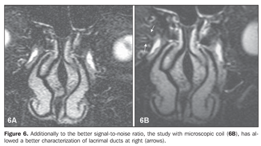
In 36 of the 40 (90%) structures evaluated, a mean 1.17 point increase was observed in the score assigned to the images in the study of each anatomical structure with the microscopic coil as compared with the C1 surface coil. Only 10% (four anatomical structures – two nasolacrimal ducts in case 2 and two lacrimal sacs in case 5) of the whole sample did not present any increase in the score assigned to images acquired with the microscopic coil. A subjective improvement was observed in the signal-to-noise ratio for the whole sample in the comparison between images acquired with microscopic coil and with conventional surface coil.
DISCUSSION Epiphora (abnormal overflow of tears due to obstruction of lacrimal pathways) is a frequent complaint(1) in ophthalmologic offices (about 3% of ambulatory visits). Data about level and type of obstruction play a significant role in the definition of the best therapeutic strategy to be adopted (dacryocystorhinostomy, dacryocystoplasty, nasolacrimal stent or antibiotics irrigation). Additionally to the clinical evaluation, an imaging method should be utilized for this purpose. The principles of dacryocystography with conventional radiography were first described by Ewing in 1909 and, up to recently, this was the method most frequently utilized in the evaluation of the lacrimal system. Despite providing an excellent anatomical delineation, this technique is relatively invasive and involves certain risks such as iatrogenic trauma and post-catheterization stenosis, besides the harmful effects of the ionizing radiation on the crystalline lens, without offering any functional information, considering that the contrast agent is administered under pressure by a syringe/catheter. Dacryoscintigraphy is an alternative method for evaluating functional alterations of the lacrimal pathways. However, the following disadvantages of this method should be considered: the low spatial resolution for orbital soft tissues imaging and crystalline lens exposure to radiation. Despite representing an important alternative in the study of lacrimal pathways, computed tomography dacryocystography also presents relative disadvantages connected with the utilization of ionizing radiation and the potential conjunctival irritation caused by the iodinated contrast agent. MRI has been reported in the literature as a feasible method for evaluating the lacrimal pathways. The main advantages in relation to the other methods are: non-invasiveness, absence of ionizing radiation, (instillation rather than catheterization), anatomical and functional evaluation, and feasibility of simultaneous bilateral evaluation. Either T1- or T2-weighted sequences can be utilized for images acquisition(2–7). In the first case, a gadolinium solution acts as a contrast agent; on T2-weighted sequences, likewise in the present study, the 0.9% saline solution is the medium that allows the identification of the lacrimal pathways.
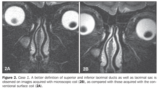
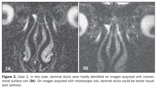
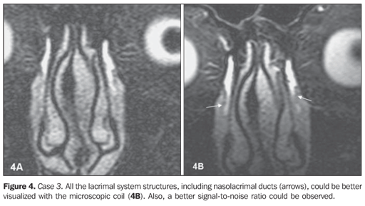
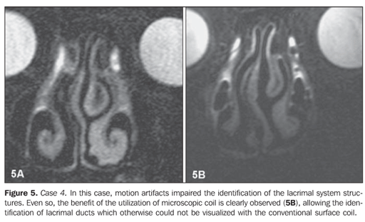
Surface coils allow high-quality imaging of superficial anatomical structures like the normal lacrimal pathways structures. Conventional surface coils, with larger diameters, can provide images with a non-optimum sinal-to-noise ratio, utilizing very thin slices. In the present protocol, 1.8 mm-thick slices presented a poor signal-to-noise ratio with the conventional coil. Despite the fact that the majority of structures could be visualized with this coil, their visualization was rated as non-optimum. Several studies in the literature report an improvement of the signal-to-noise ratio with the utilization of microscopic coils for evaluating several structures of different tissues such as knee ligaments and menisci(8) and pathological cervical lymph nodes(9). The present study is compatible with these reports, considering that all the lacrimal system structures were better characterized in the evaluation with the microscopic coil, even in the presence of artifacts (motion artifacts, for example). This aspect is particularly relevant if the identification of lacrimal ducts is considered. Considering their small caliber, sometimes the visualization of these structures with conventional surface coils is limited(2). In the present study, the lacrimal ducts characterization improved with the utilization of the microscopic coil. This finding allows the authors to consider the possibility of improvement in the diagnosis of lacrimal pathways obstruction, especially high (pre-saccular) obstructions. However, further studies with symptomatic individuals would be necessary to confirm this hypothesis.
CONCLUSION Magnetic resonance imaging dacryocystography utilizing microscopic coils is the appropriate method for identifying normal structures of the lacrimal pathways, resulting in higher-quality images as compared with images acquired with conventional surface coils.
REFERENCES 1. Linberg JV, McCormick SA. Primary acquired nasolacrimal duct obstruction. A clinicopathologic report and biopsy technique. Ophthalmology. 1986;93:1055–63. [ ] 2. Caldemeyer KS, Stockberger SM Jr, Broderick LS. Topical contrast-enhanced CT and MR dacryocystography: imaging the lacrimal drainage apparatus of healthy volunteers. AJR Am J Roentgenol. 1998;171:1501–4. [ ] 3. Kirchhof K, Hähnel S, Jansen O, et al. Gadolinium-enhanced magnetic resonance dacryocystography in patients with epiphora. J Comput Assist Tomogr. 2000;24:327–31. [ ] 4. Yoshikawa T, Hirota S, Sugimura K. Topical contrast-enhanced magnetic resonance dacryocystography. Radiat Med. 2000;18:355–62. [ ] 5. Takehara Y, Isoda H, Kurihashi K, et al. Dynamic MR dacryocystography: a new method for evaluating nasolacrimal duct obstructions. AJR Am J Roentgenol. 2000;175:469–73. [ ] 6. Manfrè L, de Maria M, Todaro E, et al. MR dacryocystography: comparison with dacryocystography and CT dacryocystography. AJNR Am J Neuroradiol. 2000;21:1145–50. [ ] 7. Hoffmann KT, Hosten N, Anders N, et al. High-resolution conjunctival contrast-enhanced MRI dacryocystography. Neuroradiology. 1999;41: 208–13. [ ] 8. Niitsu M, Ikeda K. Magnetic resonance microscopic images with 50-mm field-of-view of the medial aspect of the knee. Acta Radiol. 2004;45: 760–8. [ ] 9. Sumi M, Van Cauteren M, Nakamura T. MR microimaging of benign and malignant nodes in the neck. AJR Am J Roentgenol. 2006;186:749–57. [ ] Received September 4, 2007. Accepted after revision December 12, 2007. * Study developed at Scopo Diagnóstico, Unit of US, CT and MRI, Hospital São Luiz, São Paulo, SP, Brazil. |
|


Av. Paulista, 37 - 7° andar - Conj. 71 - CEP 01311-902 - São Paulo - SP - Brazil - Phone: (11) 3372-4544 - Fax: (11) 3372-4554