Radiologia Brasileira - Publicação Científica Oficial do Colégio Brasileiro de Radiologia
AMB - Associação Médica Brasileira CNA - Comissão Nacional de Acreditação
 Vol. 41 nº 4 - July / Aug. of 2008
|
ORIGINAL ARTICLE
|
|
Radiographic analysis of the cervical spine in healthy individuals submitted to manual traction |
|
|
Autho(rs): Roger Burgo de Souza, Edson Lopes Lavado, Fausto Orsi Medola, Dirceu Henrique Blanco, João Henrique Blanco |
|
|
Keywords: Radiography, Cervical spine manipulation, Muscle stretching exercise |
|
|
Abstract:
IMaster, Professor, Universidade Estadual de Londrina (UEL), Londrina, PR, Brazil
INTRODUCTION The cervical spine consists of seven vertebrae constituting a structure connecting the head to the trunk, with the function of supporting the skull, maintaining the alignment and enabling head motion(1). This pillar of movable bones is supported by ligamentous structures constituting a passive stability system(2). This region dynamics is under the control of cervical muscle groups, and adjacent to this complex of osteomyoligamentous structures there are cartilaginous, nervous, glandular tissues and arteriovenous components(3). Manual therapy encompasses manipulation, passive mobilization, neuromuscular therapy, manual traction, soft tissue massage(4,5), utilized in the physiotherapy practice as a complementary procedure in the reduction of algesic processes and osteomyoneuroarticular alterations of the cervical and lumbar regions. The manual cervical traction technique consists of a separation force applied by a physiotherapist's hands to separate the vertebrae(6), and is considered as a therapeutic modality that can be administered with the patient in the supine or sitting position(7,8). Countless biomechanical studies have tried to determine the mechanism employed in the cervical traction technique, including vertebral motion, intervertebral foramen separation, the best traction angle, the application of appropriate load (force), the ideal traction duration and ligamentous deformation(5,8). Although cervical traction is widely utilized in the treatment of several types of compressive and tensional cervical disorders, opinions about methods of application and clinical results are divergent(9), because several studies fail in demonstrating the relation between timing and load magnitude, and conservative treatments still lack standardization(10). However, the advantages of manual traction include easy hands positioning, sensory feedback, technique specificity and comfort of the patient (considering that the technique is applied with the patient at rest). Some physiological effects of the manual traction include decompression of articular, neurological and vascular structures; soft tissue lengthening and mechanoreceptors stimulation to provide pain relief and reduction of the muscular tonus(11). There are different forms of applying cervical traction techniques, ranging from manual cervical traction to other types of mechanic traction - intermittent and static -, which makes a comparison among studies more difficult(12). Additionally, there is a scarcity of studies in the literature reporting the measurement of the length of the cervical spine under manual traction. Radiological evaluation is considered as a golden standard, allowing the evaluation of the cervical range of motion besides its morphology(13). Some researchers have utilized cervical radiology to measure the vertebral angle, separation and motion by means of mechanical devices(14,15) . The present study is aimed at radiographically evaluate asymptomatic individuals submitted to manual traction for changes in the cervical spine length between the second (C2) and seventh (C7) vertebrae.
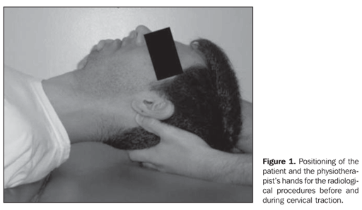
MATERIALS AND METHODS The individuals were invited to participate in the study, constituting a convenience sampling with 55 male and female individuals in the age range between 19 and 25 years. Individuals with previous history of cervical conditions, traumatisms (recent or not), postural alterations, suspected pregnancy, or utilizing myorelaxant drugs were not included in the present study. All the participants were given an explanation about the procedures to be adopted during the experiment as well as the phases of the study, and signed a term of free and informed consent approved by the local Committee for Ethics in Research, according to the Resolution Nbr. 196/96 of the Brazilian Health Council/Ministry of Health. Experimental procedure All the individuals were evaluated for age, sex and body mass. In order to minimize errors in positioning or in the technique execution, all the participants were previously familiarized with manual cervical traction. Radiological examinations and manual traction were pre-scheduled for times in the morning period. The identification of the radiographic films was based on a random number table to allow a blind evaluation both before and during the manual traction. Initially, the patients were positioned in dorsal decubitus on the examination table of a DC-15KB, 500 mA X-ray equipment (Toshiba; Tokyo, Japan), with the cervical region without clothing and without any metal ornament that could interfere with the technique execution. The cervical spine was positioned with the chin at a 90º angle, confirmed by means of a large, transparent plastic goniometer scale with two measuring rules and a 0° - 360° protractor (Carci; São Paulo, Brazil). The patients were instructed to depress their shoulders, allowing an appropriate visualization of the neck, and maintaining a static and relaxed posture, after confirmation of the positioning by the goniometer, until the whole radiological procedure with manual cervical traction was completed. Subsequently, the physiotherapist stayed at the upper end of the examination table with his right hand fixing the patient's mandible body, and the left hand positioned under the occipital region, with the first and third fingers touching the mastoid process at each side of the patient's skull. Then, the radiology technician positioned the X-ray tube calibrated for 50 kV penetration and 0.8 mAs radiation time, directed to the center of the fourth cervical vertebra, laterally to the neck, at a 80 cm distance for obtention of a lateral view on a plain, standard 24 cm × 30 cm radiographic film (initial radiography). Following the confirmation by the radiology technician that the radiographic film met the criteria for a correct visualization of all cervical vertebrae, the therapist started the manual traction, applying a longitudinal separation force between the head and the trunk during 120 seconds. Another X-ray beam was shot for obtaining the second radiographic image (final radiography) (Figure 1). In case of failure in the film processing, in the equipment performance or postural change, a repetition of the procedures was scheduled for 30 days later.
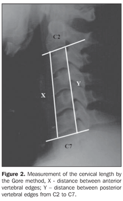
Procedure for radiographic films measurement One hundred and ten radiographic images obtained before and during the manual cervical traction were randomly numbered (0 to 110) with the aid of a random number table to allow a blind evaluation by the observer. The films were placed in envelopes duly identified with the name of the research project which were sent to the radiologist for measurement of the C2-C7 distances. For these measurements the radiologist utilized the Gore method(16) , drawing a tangent to the lower edge of the C2 vertebral body and another on the up-per edge of the C7 vertebral body. Once these tangent lines were drawn on plain radiographs with lateral views, the distances between the anterior vertices and between the posterior vertices of the respective vertebrae were measured in centimeters (Figure 2).
Along all the phases of the present study, the physiotherapist, the radiology technician and the radiologist were the same to avoid any bias. Statistical analysis The descriptive statistics expressed mean values and standard deviation or median, quartiles and minimum and maximum values presented by means of Figures and Tables. The Student's t test was utilized for comparison age, body mass, height and body mass index variables. The Wilcoxon test was utilized in the comparison between findings before and during manual cervical traction. The Statistical Package for Social Sciences software, version 13.0 was utilized. The level of statistical significance of all the mentioned tests was established in 5% (p = 0.05).
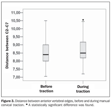
RESULTS Twelve (22%) of the 55 individuals were men and 43 (78%) women. Mean age was 21.20 ± 1.40 years, mean body mass, 58.40 ± 9.70 kg, mean height, 1.66 ± 0.08 m, and the mean body mass index, 20.96 ± 2.14 kg/m². As regards the length of the cervical spine in the comparison between the anterior vertebral edges before the traction, the median was 8.40 cm (minimum = 7.05 cm, maximum = 10.50 cm, 1st quartile = 8.00 cm, 3rd quartile = 9.00 cm) and during traction was 8.50 cm (minimum = 7.20 cm, maximum = 10.90 cm, 1st quartile = 8.20 cm, 3rd quartile = 9.20 cm). The comparison between anterior vertebral edges values before and during traction resulted in a statistically significant difference (p = 0.002) (Figure 3).
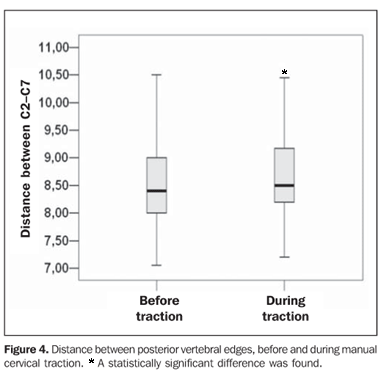
In the measurements between the posterior vertebral edges before the traction, the median was 8.35 cm (minimum = 7.10 cm, maximum = 10.30 cm, 1st quartile = 7.95 cm, 3rd quartile = 8.70 cm) and during traction was 8.50 cm (minimum = 7.30 cm, maximum = 10.50 cm, 1st quartile = 8.00 cm, 3rd quartile = 8.90 cm). The comparison between posterior vertebral edges values before and during traction resulted in a statistically significant difference (p < 0.001) (Figure 4).
DISCUSSION The measurement of the cervical spine length by means of radiography demonstrated to be simple, low-cost procedure, and easy to perform. The choice of the imaging method was based on the cost/benefit ratio for the present study, considering that the radiological evaluation is considered as the golden-standard, enabling the radiographic evaluation of the angular and linear limits of the cervical spine motion, besides its morphometry(13,17). Although magnetic resonance imaging is the best method for demonstrating tissues differences and conditions, this method is still expansive, time consuming and many times is not readily available(18). In a study developed by Vaughn et al.(14), a radiographic analysis of asymptomatic individuals was performed to investigate the angle and increase in the intervertebral space with mechanical traction or pre-established loads. In the present study, the results demonstrated that the strength applied by the physiotherapist increased the length of the cervical spine between the C2 and C7 vertebrae measured by means of plain lateral radiographic views of asymptomatic individuals. It can be concluded that manual traction results in an increase of intervertebral spaces and relaxation of muscular structures, similarly to the results of previous studies with the utilization of mechanical traction(8,17,19). Although cervical traction is widely employed in the treatment of several compressive and tensional cervical disorders, opinions diverge about application method and clinical results(9), because several studies fail in demonstrating the relation between timing and load magnitude, and conservative treatments still lack standardization(10). However, in the present study, there was a concern regarding the supine positioning of the individual in order to avoid any inappropriate posture, maintaining a neutral cervical spine positioning, allowing a single load vector, and timing (120 seconds) previously established according to the time achieved by the physiotherapist while sustaining the traction force. It is understood that this traction technique is employed to achieve a stretching of cervical muscles, as well as an increase of intervertebral spaces which under tension and narrowed constitute respectively the etiological factors in algesic and compressive processes of this region(7). The mechanical cervical traction effect is a decrease in the deficit of palmar pressure in individuals with cervical radiculopathies(8). The advantages of manual traction as compared with mechanical traction include easy hands positioning, sensory feedback, technique specificity and comfort of the patient (considering that the technique is applied with the patient at rest). Some physiological effects of the manual traction include decompression of articular, neurological and vascular structures; soft tissue lengthening and mechanoreceptors stimulation to provide pain relief and reduction of the muscular tonus(11). However, the knowledge of medical diagnosis besides a functional kinesiological evaluation is necessary aiming at an appropriate therapy planning for the management of cervical disorders. In another study, patients diagnosed with radiculopathy with herniation > 4 mm have been submitted to intermittent cervical traction and evaluated by magnetic resonance imaging. The traction was sustained for 45 minutes during six to eight hours with pre-established loads. All the patients have presented an improvement in symptoms and disc herniation reduction(19). Notwithstanding the distances between each vertebral body have not been measured in the present study, it can be concluded that the longitudinal force applied aids in the reduction of the pressure between vertebral bodies, resulting in an increase of intervertebral spaces and, consequently in the length of the cervical spine. Chung et al.(17) studied an asymptomatic group and another with diagnosis of cervical disk protrusion, in which intermittent cervical traction was applied during daily life activities by means of a pneumatic device calibrated in 30-pound strength. Subsequently, the individuals were submitted to magnetic resonance imaging that demonstrated an increase in the cervical spine length. In the asymptomatic group, a 1.93 mm increase was observed, and in the group with hernia, 2.19 mm, with a significant decrease in the disk protrusion. These results were similar to the findings of the present study, confirming again that the cervical traction affects the length of the cervical region, and that manual traction and mechanical traction produce similar alterations. A study involving cervical spines of cadavers submitted to mechanical traction associated with cervical spine flexion has observed a 3–4 mm² increase in the intervertebral foramen(20). However, these results are not compatible with those in living individuals. The foramen was not investigated in the present study, but it is believed that the summation of these values would result in an increase in the longitudinal length of the cervical spine of living individuals. In contrast to the results of the present study, other study(5) has found a decrease of approximately 5 mm in the length between the posterior edges of the vertebral bodies from C2 to C7 during traction associated with mechanical of the cervical region. It is considered that this association has fostered an increase in the cervical lordosis, resulting in a reduction of the posterior length of the vertebral bodies. In the present study, there was a concern regarding the supine positioning of the individual, in order to avoid compensation for the cervical curvature. There are evidences that facets sliding occur between cervical vertebrae at the cervical spine extension, contributing to an approximation of the posterior vertebral bodies(1). Along all the phases of the present study, none of the individuals reported discomfort or pain during or after the manual cervical traction. Notwithstanding the achievement of a potentially beneficial alteration of the cervical spine by means of manual cervical traction, the authors of the present study were faced with a limitation concerning the degree of manual force required to increase the intervertebral spaces, considering that previous studies reported procedures of cervical traction with predefined loads(8,20). The authors believe that another limitation was the fact of the convenience sample being constituted by asymptomatic individuals, not allowing an evaluation of symptoms that could emerge or decrease before, during or after the manual traction. Another limitation was the age range (19–25 years) that is not appropriately representative of the adult population with cervical dysfunctions. The present study demonstrated that manual cervical traction increases the length of the cervical spine, fostering an increase in the distance between vertebral bodies, radicular decompression and cervical muscles relaxation, and should be a complement to the therapeutic procedures included in a physiotherapy program.
CONCLUSION Based on the comparison between measurements of the cervical spine before and during manual cervical traction, the present study demonstrated satisfactory outcomes, with a statistically significant increase in the length of the cervical spine. Specific therapeutic exercises, such as manual cervical traction should be a key factor in a better prognosis for osteomyoneuroarticular involvement of the cervical spine. Therefore, the radiological analysis will play a significant role in the interpretation and demonstration of the benefits of this physiotherapeutic intervention. However, further random clinical studies are required for defining an effective therapeutic strategy aiming at adjusting the cervical length as well as calculating the appropriate manual strength to be applied in these procedures in asymptomatic individuals. Acknowledgements The sincere thanks of the authors are due to the Unit of Radiology, Hospital Universitário, Mr. Jaime Stelli, Radiology Technician, for his availability and scheduling of radiological procedures, and the Clinical Board, represented by Dr. Sinésio Moreira Junior, for authorizing and supporting the development of this project, besides those who voluntarily participated in the present study.
REFERENCES 1. Mercer SR, Bogduk N. Joints of the cervical vertebral column. J Orthop Sports Phys Ther. 2001; 31:174–82. [ ] 2. Olson KA, Joder D. Diagnosis and treatment of cervical spine clinical instability. J Orthop Sports Phys Ther. 2001;31:194–206. [ ] 3. Nordin M, Frankel VH. Biomecânica da coluna cervical. In: Nordin M, Frankel VH, editores. Biomecânica básica do sistema musculoesquelético. 3ª ed. Rio de Janeiro: Guanabara Koogan; 2003. p. 250–60. [ ] 4. Gross AR, Kay T, Hondras M, et al. Manual therapy for mechanical neck disorders: a systematic review. Man Ther. 2002;7:131–49. [ ] 5. Harrison DE, Cailliet R, Harrison DD, et al. A new 3-point bending traction method for restoring cervical lordosis and cervical manipulation: a nonrandomized clinical controlled trial. Arch Phys Med Rehabil. 2002;83:447–53. [ ] 6. Carette S, Fehlings MG. Cervical radiculopathy. N Engl J Med. 2005;353:392–9. [ ] 7. Nadler SF. Nonpharmacologic management of pain. J Am Osteopath Assoc. 2004;104(11 Suppl 8):S7–12. [ ] 8. Joghataei MT, Arab AM, Khaksar H. The effect of cervical traction combined with conventional therapy on grip strength on patients with cervical radiculopathy. Clin Rehabil. 2004;18:879–87. [ ] 9. Wong AMK, Lee MY, Chang WH, et al. Clinical trial of a cervical traction modality with electromyographic biofeedback. Am J Phys Med Rehabil. 1997;76:19–25. [ ] 10. Rodacki ALF, Weidle CM, Fowler NE, et al. Changes in stature during and after spinal traction in young male subjects. Rev Bras Fisioter. 2007;11:63–71. [ ] 11. Makofsky HW. Técnicas para os tecidos conjuntivos e procedimentos de alongamentos para a coluna cervical. In: Makofsky HW, editor. Coluna vertebral: terapia manual. 1ª ed. Rio de Janeiro: Guanabara Koogan; 2006. p. 90–101. [ ] 12. Peake N, Harte A. The effectiveness of cervical traction. Physical Therapy Reviews. 2005;10: 217–29. [ ] 13. Wolfenberger VA, Bui Q, Batenchuk GB. A comparison of methods of evaluating cervical range of motion. J Manipulative Physiol Ther. 2002;25: 154–60. [ ] 14. Vaughn HT, Having KM, Rogers JL. Radiographic analysis of intervertebral separation with a 0º and 30º rope angle using the Saunders cervical traction device. Spine. 2006;31:39–43. [ ] 15. Fernández-de-las-Peñas C, Downey C, Miangolarra-Page JC. Immediate changes in radiographically determined lateral flexion range of motion following a single cervical HVLA manipulation in patients presenting with mechanical neck pain: a case series. Int J Osteopath Med. 2005;8:139–45. [ ] 16. Rowe LJ, Yochum TR. Measurements in skeletal radiology. In: Rowe LJ, Yochum TR, editors. Essential of skeletal radiology. 2nd ed. New York: Williams & Wilkins; 1999. p. 153–4. [ ] 17. Chung TS, Lee YJ, Kang SW, et al. Reducibility of cervical disk herniation: evaluation at MR imaging during cervical traction with a nonmagnetic traction device. Radiology. 2002;225:895–900. [ ] 18. Nucci CG, Caiero MT, Lasmar RCP, et al. Estudo radiográfico da coluna cervical em crianças com artrite reumatóide juvenil. Rev Bras Ortop. 1999; 34:117–27. [ ] 19. Constantoyannis C, Konstantinou D, Kourtopoulos H, et al. Intermittent cervical traction for cervical radiculopathy caused by large-volume herniated disks. J Manipulative Physiol Ther. 2002;25:188–92. [ ] 20. Humphreys SC, Chase J, Patwardhan A, et al. Flexion and traction effect on C5-C6 foraminal space. Arch Phys Med Rehabil. 1998;79:1105–9. [ ] Received September 20, 2007. Accepted after revision December 14, 2007. * Study developed at Universidade Estadual de Londrina (UEL), Londrina, PR, Brazil. |
|
Av. Paulista, 37 - 7° andar - Conj. 71 - CEP 01311-902 - São Paulo - SP - Brazil - Phone: (11) 3372-4544 - Fax: (11) 3372-4554


