Radiologia Brasileira - Publicação Científica Oficial do Colégio Brasileiro de Radiologia
AMB - Associação Médica Brasileira CNA - Comissão Nacional de Acreditação
 Vol. 41 nº 2 - Mar. / Apr. of 2008
|
ORIGINAL ARTICLE
|
|
Impact of a program of diagnostic imaging quality control in mammography centers of the Federal District, Brazil |
|
|
Autho(rs): Rosangela da Silveira Corrêa, João Emílio Peixoto, Lynn Dee Silver, Cintia Melazo Dias, Maria do Socorro Nogueira, Suy Ferreira Hwang, Rubemar de Souza Ferreira |
|
|
Keywords: Mammography, Quality control, Federal District, Impact |
|
|
Abstract:
IMaster, Senior Technologist, Centro Regional de Ciências Nucleares do Centro–Oeste – Comissão Nacional de Energia Nuclear (CRCN–CO/CNEN), Abadia de Goiás, GO, Brazil
INTRODUCTION Two types of cancer stand out amongst women: uterine cervix cancer and breast cancer. The first one is the main cause for cancer deaths in the Center–Western and Northern regions, while breast cancer, in the other regions of the country. According to data from the Ministry of Health, breast cancer kills more Brazilian women in than any other type of cancer. Breast cancer, among the 237,480 new cases estimated for 2006, would be the main type of neoplasm to affect the female population, possibly accounting for up to 48,930 new cases. The Federal District presents an estimated gross incidence of 53 cases for each 100,000 women(1). In the absence of effective primary prevention mechanisms efforts should be focused on the early detection of this disease. Mammography is a method utilized for detection and diagnosis of breast diseases, and frequently is performed in healthy women who intend to maintain this status(2). However, this type of diagnostic method presents some limitations, and the screening may result in adverse consequences. A frequent problem is the false–positive result, where the suspicion for a malignant lesion is not confirmed after histopathological studies. Another limitation is represented by false–negative results that may lead to the postponement of an appropriate action in relation breast cancer. Taking these problems into consideration, the development of quality control programs represents a necessity in terms of efficiency, and an obligation in ethical and moral terms(3). The imaging quality reduces, although does not eliminate, the occurrence of false–positive and false–negative results. Therefore, all mammography centers must be focused on a permanent improvement of their services in order to achieve a harmonious integration among the following areas: medical, technological, administrative, economic, public health assistance, and, if applicable, academic and research. The greatest risk for a woman who has been submitted to mammography is that a small breast cancer goes undetected because of the low quality of mammographic images. This risk is ten times higher than the risk for a radiation–induced breast cancer(4,5). The present comparative study was developed in mammography centers in the Federal District, Brazil, over the period from 2000 to 2002, including a chronological results (before and after–type analysis).
MATERIALS AND METHODS The term "intervention", in the present study, is defined as a sequence of actions involving: inspection (first and second technical visits), qualification courses for both radiologists and technicians, and notification from the Departamento de Fiscalização de Saúde do Distrito Federal (DpFS–DF) (Federal District Department of Health Inspection) to mammography centers requesting irregularities correction. The results evaluation was focused on the quality of images for early breast cancer detection in compliance with the technical requirements included in the Order (Portaria) no. 453(6) of the Brazilian Ministry of Health, the European guidelines for quality assurance in mammography screening(7) and the American College of Radiology guidelines for breast cancer screening(8). The performance of the whole mammograms production chain in mammography centers was quantitatively evaluated with the aid of a breast phantom comprised of test structures in several shapes, sizes and compositions, and producing images similar to the anatomical structures of interest, besides an optical densities scale. The radiologic breast phantom was developed by the Unit of Radiology at Santa Casa da Misericórdia do Rio de Janeiro(9) and approved by Colégio Brasileiro de Radiologia (Brazilian College of Radiology)(10). The evaluation included the following items: a) Alignment between the X–ray field and the images receptor; b) automatic exposure control performance testing; c) breast compression rate; d) alignment of the breast compression paddle; e) evaluation of the images recording system (cassette holders – screen–film contact); f) films processing quality; g) imaging quality: –images definition (spatial resolution); –high–contrast detail; –low–contrast threshold; –low–contrast linear detail; –tumor–like masses; –background optical density.
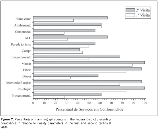
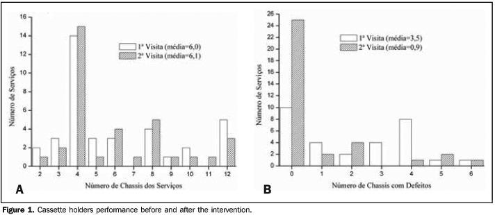
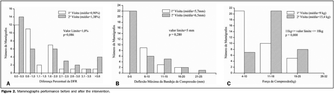
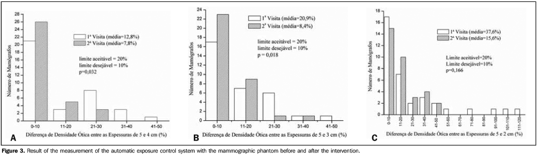
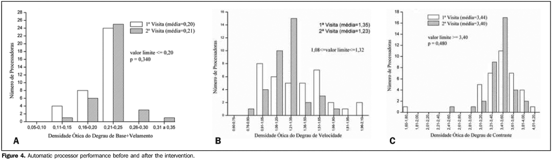
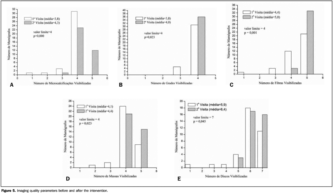
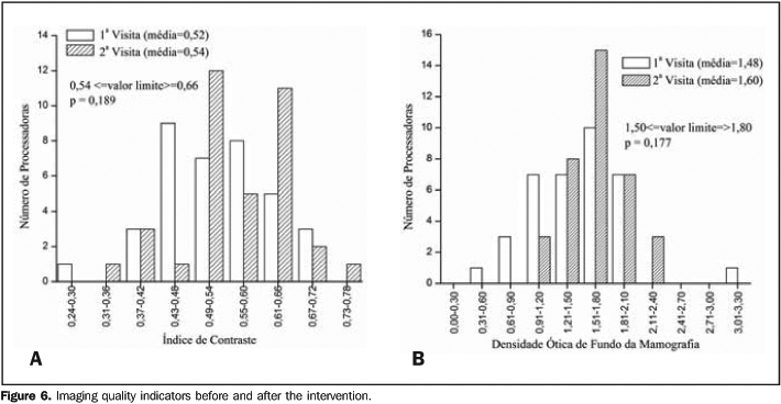
RESULTS Forty–one mammography centers in the Federal District were enrolled in the present study. However, only 36 of them were evaluated, considering that five units were excluded from the study right away at the first visit, for not meeting the inclusion criteria. This fact reflects the significance of the impact at the first phase of the intervention, considering that three of these centers (7.3%) were closed down because of quality–related issues. Evaluation of imaging quality in mammography centers of the Federal District Imaging quality indicators are gathered in two different sets of results: the first one includes technical and performance parameters of mammographs and processors; the second one includes parameters regarding the final quality of the images from a radiologic breast phantom recorded on a radiographic film. Figure 1A shows the distribution of cassette holders utilized in the mammography centers. It may be observed that this number has not changed between the first and second technical visits. On the other hand, Figure 1B clearly demonstrates that the number of defective cassette holders in each center changed significantly, especially regarding the number of centers with no defective cassette holder that changed from 10 at the first visit, to 25 at the second. As regards the performance of the mammographs technical parameters evaluated in the two visits, Figure 2A demonstrates that the results from the measurements of alignment between X–ray field and the chest wall of the patients did not present a statistically significant improvement (p = 0.086). The same occurred in relation to the measurement of maximum compression paddle deflection, with a non–statistically significant difference (p = 0.280) between the two technical visits, as shown on Figure 2B. The third parameter evaluated — breast compression rate — presented a statistically significant difference (p = 0.000) between the two evaluations. Figure 2C demonstrates that the number of mammographs in compliance with this parameter increased from seven to 20 between the first and the second evaluations. The appropriate operation of the automatic exposure control system is essential in the practice of mammography and constitutes a feature of the images production chain evaluated in the present study. Figure 3 shows the results from measurements of the differences among optical densities for different breast thicknesses (5 cm, 4 cm, 3 cm and 2 cm). Additionally, Figure 3 demonstrates a statistically significant improvement in the operation of automatic exposure control systems, most appropriately balancing the images darkening effect for 4 cm and 3 cm–thick breasts. However, no improvement was observed in the performance of this device for imaging 2 cm–thick breasts. The performance of automatic films processors was evaluated by means of a sensitometric test measuring the optical densities at three different points on a gray scale with 21 optical steps. Measurement points were the following: the base+fog step, the speed step, and the contrast step. Figure 4A demonstrates that the base density plus fog of mammograms, both in the first and second technical visits, was within the range recommended for this parameter in the greatest majority of centers, with no statistically significant difference (p = 0.340) between the two data set regarding this parameter. As regards the measurement of the speed step optical density, Figure 4B demonstrates a statistically significant variation (p = 0.034) between the first and second evaluations, and an increase in the frequency, from six to 15, of the number of films processors operating within the range recommended for this parameter. For the contrast step optical density, Figure 4C shows that not only the difference between the two data sets was not statistically significant (p = 0.480), but also there was no increase in the number of processors in compliance with the reference limit for this parameter. As regards the evaluation of final quality parameters for the images of a radiologic breast phantom recorded on the radiographic film, Figure 5 shows the results for visualization of relevant imaging details in mammography. This figure demonstrates that, for microcalcifications, masses, metal grids, fibers, and low–contrast disks, the results presented a statistically significant improvement between the first and second evaluations. Additionally, as regards other mammograms quality indicators, Figure 6 demonstrates that, although the results both for the image contrast index and background optical density have not presented a statistically significant difference (p > 0.05), there was an increase in the frequency of processors in compliance with the ranges recommended for these parameters. Considering the quality tests applied, Figure 7 shows the percentage of mammography centers in compliance with each of the parameters evaluated. As regards radiographic films processing, even with the increase (from 28% to 61%) in the number of compliant centers, this still remains as a critical issue in the mammographic images production chain. This is confirmed by the visualization of low–contrast disks that, by the time of the first technical visit was compliant in 33% of the centers, and in the second visit was compliant in 58%. This is the evidence that the reference limit for visualization of these testing objects is the most reliable imaging quality indicator reflecting the quality in the radiographic films processing.
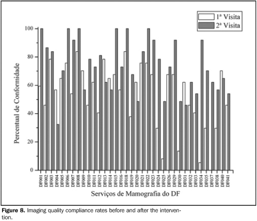
Other results regarding imaging quality indicators show that metal grids and microcalcifications imaging is poorly or not affected at all by the processing, considering their nature (high–contrast); and, also, that images of fibers and tumor–like masses relatively thick in terms of mammography are poorly affected by the processing. As regards these four imaging quality indicators, the percentage of compliant mammography centers increased from about 80% in the first technical visit to about 100% in the second one. Results regarding mammographs performance and evaluation of the images recording systems indicative of the operational status of the radiographic cassette holder (screen/film contact) show that, despite the improvement from the first to the second technical visit, the percentage of compliant centers in these items remained below or around 60%. Intervention impact on mammography centers Figure 8 shows the compliance rate resulting from the tests described in the present study. In the evaluation of the intervention impact on the 36 mammography centers of the Federal District, the mean rate of compliance regarding imaging quality increased from 53.61% to 73.19% between the first and second technical visits. According to the paired t test, at 5% significance level and with 35 freedom degrees, the difference between means was statistically significant, with the intervention affecting the improvement of the imaging quality in mammography centers.
DISCUSSION The performance of mammographs operational parameters most directly related to the imaging quality was evaluated by means of the previously mentioned tests. It should be emphasized that these tests were based on the requirements clearly established by the Order (Portaria) no. MS 453/98. The most significant results for mammographs performance in the present study were those regarding automatic exposure control systems. Figure 8 shows that, despite de increase in the percentage of compliant centers in this item, from 30% in the first technical visit to 66% in the second one, several mammography devices in the Federal District still require technical adjustments. The higher disagreement was observed in the difference between optical densities of 2 cm–thick breasts and 5 cm–thick breasts. This same situation is reported in the literature. LaFrance et al.(11), discussing similar results, have concluded that the inappropriate performance of the automatic exposure control system may be associated with the change in the X–ray beam energies spectrum caused by breast tissues attenuation (beam hardening), failure in reciprocity (to maintain a constant darkening for increasingly shorter exposure times) of the radiographic film response or to a current generated in the circuit itself in the absence of radiation exposure. According to the authors, the prevalent effect is the beam hardening. This effect may be explained as follows: as the compressed breast thickness increases, the emerging X–ray beam penetration increases, and a high percentage of photons is transmitted through the intensifier screen and the radiographic film, falling upon the sensor of the automatic compensation device. In summary, the higher the breast thickness, the greater is the beam hardening and the larger is the amount of energy absorbed by the sensor in relation to the energy absorbed by the cassette holders. Therefore, the film darkening increases as the breast thickness decreases, and vice–versa. According to the above arguments, and considering the results reported in the present study, the key issue in the performance of the automatic exposure control system is the maintenance of the film darkening between the 2 cm and 5 cm compressed breast thicknesses. It is important to emphasize that the radiologic breast phantom utilized in the present study demonstrated to be appropriate for evaluating the performance of this technical feature in mammographs. This appropriateness is due to the fact that this phantom is constituted by layers simulating breasts of different thicknesses. The other technical features of mammographs performance evaluated in the present study were the following: alignment between the X–ray field and the patient´s chest wall, the maximum compression paddle deflection, and the breast compression rate. Results presented on Figures 3A,B and 8 indicate that the percentage of compliant centers regarding alignment between field and compression paddle has not changed between the first and second technical visits, remaining in the range between 50% and 60%. The influence of the non–compliance on these two technical features could not be estimated, considering that the radiologic breast phantom utilized did not require compression to be imaged, so the phantom image quality was not affected by the compression paddle. This also is valid for the breast compression rate. Although Figure 3C demonstrates that the number of compliant mammographs in relation to this parameter increased from seven units in the first technical visit to20 units in the second visit, the phantom image could not demonstrate this improvement in the mammograph performance. Based on the above considerations, it is clear that the phantom image should not be the sole element to be considered in the evaluation of mammographic images quality, considering that several mammographs performance features related to breast compression and positioning cannot be evaluated by means of phantom images. As regards the processors performance evaluation, the present study demonstrated that, although the percentage of compliant centers has increased from 28% in the first technical visit to 61% in the second visit, this is the main source of images quality loss. Similar results can be found in the literature. Hendrick et al.(12) have reported that about 47% of mammography centers lacking approval by the American College of Radiology Accreditation Program presented non–compliance regarding processors performance. Previously, Galkin et al.(13) had already reported similar results. Approximately 40% of the films processors evaluated presented an excessive variation in their performance over a 15–day period. According to the authors, this was the main reason for the variation in the images quality and radiation doses among the facilities participating in a regional program developed in the United States for early detection of breast cancer.
CONCLUSION After an intervention process in mammography centers of the Federal District, the impact on the imaging quality, although positive both in terms of quality and in terms of quantity, has shown to be beneath the desired target–conformity level of > 90%. Several factors may have contributed for this partial result, among others, the unsatisfactory performance of films processors, poorly adjusted devices for controlling exposure and other operational parameters of the mammograph. The results from the intervention performed in the present study were similar to the ones achieved in interventions performed in other Brazilian states. And the experience acquired through other sanitary vigilance programs in mammography centers, particularly the one developed in the state of Paraíba(14), shows that a compliance rate in the range of 90% only can be achieved by means a continued action.
REFERENCES 1. Estimativas da incidência e mortalidade por câncer no Brasil, 2006. Instituto Nacional do Câncer (INCA). [Acessado em 11 de abril de 2006]. Disponível em: http://www.inca.gov.br/estimativa2006 [ ] 2. Bassett LW, Hendrick RE, Bassford TL, et al. Quality determinants of mammography: clinical practice guideline no. 13. AHCPR publication no. 95–0632. Rockville: Agency for Health Care Policy and Research, Public Health Service, U.S. Department of Health and Human Services; 1994. [ ] 3. Azevedo AC, Koch HA, Canella EO. Auditoria em centro de diagnóstico mamário para detecção precoce de câncer de mama. Radiol Bras. 2005; 38:431–4. [ ] 4. Hendrick RE. Mammography quality assurance cancer. Cancer. 1993;72:1466–74. [ ] 5. Caldas FAA, Isa HLVR, Trippia AC, et al. Controle de qualidade e artefatos em mamografia. Radiol Bras. 2005;38:295–300. [ ] 6. Ministério da Saúde. Secretaria de Vigilância Sanitária. Diretrizes de proteção radiológica em radiodiagnóstico médico e odontológico. Portaria nº 453. Diário Oficial da União, 1/6/1998. [ ] 7. Perry NN, Broeders M, Wolf C, et al., editors. European guidelines for quality assurance in mammography screening. 3rd ed. Luxembourg: European Comission, Europe Against Cancer; 2001. [ ] 8. American College of Radiology. Recommended specifications for new mammography equipment. Reston: American College of Radiology; 1993. [ ] 9. Pina DR, Morceli J, Duarte SB, et al. Otimização de imagens mamográficas. Radiol Bras. 2006;39: 351–4. [ ] 10. Colégio Brasileiro de Radiologia. Boletim do CBR nº 165, novembro 2001. p. 21. [ ] 11. LaFrance R, Gelskey DE, Barnes GT. A circuit modification that improves mamographic photo–timer performance. Radiology. 1988;166:773–6. [ ] 12. Hendrick RE, Smith RA, Wilcox PA. ACR Accreditation and legislative issues mammography. In: Haus AG, Yaffe MJ, editors. Syllabus: a categorical course in physics – technical aspects of breast imaging. Oak Brook: Radiological Society of North America; 1993. p. 137–49. [ ] 13. Galkin BM, Feig SA, Muir HD. The technical quality of mammography in centers participating in a regional breast cancer awareness program. Radiographics. 1988;8:133–45. [ ] 14. Governo do Estado da Paraíba. Secretaria da Saúde. Coordenadoria de Vigilância Sanitária. Núcleo de Radiações Ionizantes. Relatório Anual de Atividades, 1999. Paraíba; 2001. [ ] Received June 27, 2007. Accepted after revision August 24, 2007. * Study developed by Centro Regional de Ciências Nucleares do Centro–Oeste – Comissão Nacional de Energia Nuclear (CRCN–CO/CNEN), Abadia de Goiás, GO, Brazil. |
|

Av. Paulista, 37 - 7° andar - Conj. 71 - CEP 01311-902 - São Paulo - SP - Brazil - Phone: (11) 3372-4544 - Fax: (11) 3372-4554