INTRODUÇÃOO carcinoma urotelial é uma neoplasia maligna rara, responsável por apenas 5% a 7% dos tumores renais e 5% dos tumores uroteliais. Embora a cirurgia radical seja considerada o tratamento padrão ouro para pacientes com carcinoma urotelial, avanços endoscópicos demonstram resultados favoráveis de tratamento e preservação renal em casos selecionados, mesmo em pacientes com rim contralateral normal
(1,2). A seleção cuidadosa de pacientes para tratamento endoscópico é crucial, portanto, a obtenção de uma amostra satisfatória para o diagnóstico é um passo essencial na determinação do tratamento e prognóstico dos pacientes portadores de carcinoma urotelial
(3).
A biópsia é um componente integral da avaliação de lesões potencialmente malignas do ureter e outras malignidades do trato urinário superior, e quando indicada é tipicamente realizada por meio de ureteroscopia. A biópsia ureteroscópica é invasiva, necessitando de anestesia geral e inserção de cateter ureteral e, portanto, pode não ser viável nos pacientes com comorbidades de alto risco. Embora a ureteroscopia continue sendo o padrão ouro, ela pode ser tecnicamente desafiadora e está associada a taxas significativas de falso-negativos, dependendo das características morfológicas da lesão
(4). Uma série de técnicas intervencionistas guiadas por exames de imagem têm sido publicadas recentemente na literatura radiológica brasileira
(5–9).
A biópsia percutânea pode ser tentada em casos selecionados em que o segmento alvo do ureter ou da pelve renal é inacessível via ureteroscopia, é predominantemente exofítico e não endoluminal, ou com resultados inconclusivos em amostras prévias.
BIÓPSIA GUIADA POR ULTRASSONOGRAFIA (TRANSABDOMINAL E ENDOCAVITÁRIA)Como método de orientação, a ultrassonografia (US) parece ter várias vantagens. Está geralmente disponível, não envolve radiação ionizante, o dispositivo é portátil e fornece imagens em tempo real (Figura 1). Infelizmente, nem todos os pequenos tumores renais podem ser visibilizados pela US, e estruturas e órgãos adjacentes não podem ser diferenciados adequadamente, como na tomo grafia computadorizada (TC). Além disso, as estruturas gasosas e ósseas podem obscurecer a visibilização
(10). No entanto, a agulha pode ser direcionada para componentes sólidos na massa e a localização ser confirmada no momento da biópsia, o que permite um posicionamento mais preciso da agulha e um melhor fragmento da lesão
(10).
 Figura 1. A:
 US da bexiga demonstrando agulha coaxial 17G × 13 cm (seta) posicionada adjacente à lesão vegetante na parede lateral esquerda da bexiga.
B: Realização da biópsia percutânea utilizando agulha tru-cut 18G × 16 cm.
C: Anatomopatológico revelou carcinoma urotelial invasivo.
A biópsia percutânea de lesões ureterais e do sistema coletor guiada por TC é uma alternativa útil para obter amostras de lesões suspeitas em situações clínicas difíceis (Figura 2). Cenários clínicos comuns apropriados para uma abordagem percutânea guiada por TC incluem pacientes com condutos urinários ileais, anatomia esfavorável, incapacidade de acessar ureteroscopicamente, impossibilidade de realização do procedimento guiado por US ou resultados de biópsia ureteroscópica negativos no cenário de alta suspeita clínica de malignidade
(11).
 Figura 2. A:
 TC de abdome, corte axial, demonstrando lesão urotelial mal definida no terço médio do rim direito.
B: Realização da biópsia (seta) guiada por TC utilizando técnica coaxial após a injeção de contraste, fase excretora.
C: Anatomopatológico revelou carcinoma urotelial invasivo.
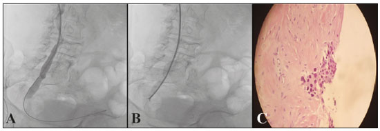
As pinças tipo fórceps demonstraram ser muito eficazes na obtenção de tecido em estenoses biliares malignas
(12,13). O material é composto por bainha e fórceps, que são avançados ao lado ou não de um fio-guia de 0,035. Esta técnica permite a biópsia da estenose por meio da bainha, deixando um fio de 0,035 mantendo o acesso através da lesão (Figura 3). Isto também é vantajoso quando a posição do fio distal é necessária para procedimentos subsequentes. Apesar de seu sucesso documentado, há poucos relatos sobre o uso e técnicas de fórceps sendo empregados para obter amostras de regiões não biliares
(14). Trata-se de uma técnica relativamente segura e eficaz, além de possibilitar o tratamento oncológico guiado por histologia
(15).
 Figura 3. A:
 Ureterografia demonstrando lesão obstrutiva no ureter distal direito e passagem do ponto obstrutivo utilizando fio-guia hidrofílico até a bexiga.
B: Bainha 7F × 45 cm posicionada até região próxima ao ponto obstrutivo e realizada biópsia com pinça tipo fórceps.
C: Anatomopatológico revelou neoplasia urotelial bem diferenciada.
As modalidades de imagem atuais fornecem a oportunidade de realização de biópsias de lesões focais em vários órgãos abdominais e pélvicos profundos, possibilitando que um diagnóstico seja feito de modo seguro e eficaz
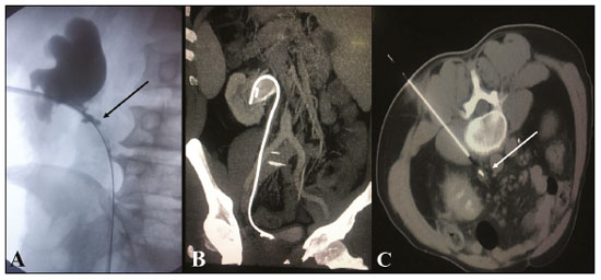
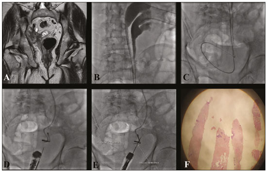
(16,17). Em situações em que os tumores da bexiga não são facilmente acessíveis para biópsia por cistoscopia, métodos alternativos para a aquisição de amostras de tecido, como TC, US ou fluoroscopia, podem ser utilizados isoladamente ou em conjunto para a realização da biópsia percutânea (Figuras 4 e 5).
 Figura 4. A:
 Pielografia pré-procedimento de inserção percutânea de cateter duplo J demonstrando estenose do ureter proximal com irregularidades parietais (seta).
B: TC de abdome coronal pós-contraste, reconstrução MIP, mostrando cateter duplo J posicionado adequadamente.
C: Biópsia percutânea guiada por TC utilizando acesso posterior e técnica coaxial demonstrando espessamento do ureter com cateter duplo J no interior para proteção ureteral. Anatomopatológico revelou pseudotumor inflamatório do ureter.
 Figura 5. A:
 Ressonância magnética de pelve ponderada em T1 demonstrando acentuada hidronefrose à direita secundária a lesão pélvica extraperitonial na região do ureter distal e com sinais de invasão da bexiga, vesícula seminal e próstata.
B: Paciente em decúbito dorsal, pielografia realizada pré-procedimento confirmando duplicação ureteral completa.
C: Inserção percutânea de cateter duplo J (cálice superior) e nefrostomia (cálice inferior).
D,E: Realizada biópsia percutânea guiada por US endorretal e fluoroscopia.
F: Relatório anatomopatológico revelou neoplasia urotelial pouco diferenciada.
A biópsia percutânea do sistema coletor do trato urinário superior guiada por imagem parece ser uma opção segura e eficaz à biópsia ureteroscópica em pacientes selecionados que falharam ou são candidatos inadequados a ureteroscopia. Em pacientes selecionados e que não possuem cateter duplo J, pode-se realizar a inserção pela técnica percutânea
(18) antes da biópsia.
REFERÊNCIAS1. Chen GL, Bagley DH. Ureteroscopic management of upper tract transitional cell carcinoma in patients with normal contralateral kidneys. J Urol. 2000;164:1173–6.
2. Elliott DS, Segura JW, Lightner D, et al. Is nephroureterectomy necessary in all cases of upper tract transitional cell carcinoma? Long-term results of conservative endourologic management of upper tract transitional cell carcinoma in individuals with a normal contralateral kidney. Urology. 2001;58:174–8.
3. Kleinmann N, Healy KA, Hubosky SG, et al. Ureteroscopic biopsy of upper tract urothelial carcinoma: comparison of basket and forceps. J Endourol. 2013;27:1450–4.
4. Vashistha V, Shabsigh A, Zynger DL. Utility and diagnostic accuracy of ureteroscopic biopsy in upper tract urothelial carcinoma. Arch Pathol Lab Med. 2013;137:400–7.
5. Schiavon LHO, Tyng CJ, Travesso DJ, et al. Computed tomography-guided percutaneous biopsy of abdominal lesions: indications, techniques, results, and complications. Radiol Bras. 2018;51:141–6.
6. Falsarella PM, Rocha RD, Rahal Junior A, et al. Minimally invasive treatment of complex collections: safety and efficacy of recombinant tissue plasminogen activator as an adjuvant to percutaneous drainage. Radiol Bras. 2018;51:231–5.
7. Tibana TK, Grubert RM, Silva CMDR, et al. Percutaneous cholangioscopy for the treatment of choledocholithiasis. Radiol Bras. 2019;52:314–5.
8. Tibana TK, Grubert RM, Santos RFT, et al. Percutaneous nephrostomy versus antegrade double-J stent placement in the treatment of malignant obstructive uropathy: a cost-effectiveness analysis from the perspective of the Brazilian public health care system. Radiol Bras. 2019;52:305–11.
9. Tibana TK, Fornazari VAV, Gutierrez Junior W, et al. What the radiologist should know about the role of interventional radiology in urology. Radiol Bras. 2019;52:331–6.
10. Lee SW, Lee MH, Yang HJ, et al. Experience ofultrasonographyguidedpercutaneous core biopsy for renal masses. Korean J Urol. 2013;54:660–5.
11. Hendrickson AC, Schmitz JJ, Kurup AN, et al. Percutaneous ureteral biopsy: safety and diagnostic yield. Abdom Radiol (NY). 2019; 44:333–6.
12. Inchingolo R, Spiliopoulos S, Nestola M, et al. Outcomes of per-cutaneous transluminal biopsy of biliary lesions using a dedicated forceps system. Acta Radiol. 2019;60:602–7.
13. Park JG, Jung GS, Yun JH, et al. Percutaneous transluminal forceps biopsy in patients suspected of having malignant biliary obstruction: factors influencing the outcomes of 271 patients. Eur Radiol. 2017;27:4291–7.
14. Carr CS, Rawlins R, Brown KM, et al. Intraluminal biopsy of a superior vena cava mass. Eur J Cardiothorac Surg. 2001;19:724–5.
15. Thomson B, Kawa B, Rabone A, et al. Fluoroscopic guided transluminal biopsy of the oesophagus and ureter with a biliary biopsy forceps kit. Cardiovasc Intervent Radiol. 2019;42:1045–7.
16. Gupta S, Madoff DC, Ahrar K, et al. CT-guided needle biopsy of deep pelvic lesions by extraperitoneal approach through iliopsoas muscle. Cardiovasc Intervent Radiol. 2003;26:534–8.
17. Gupta S, Nguyen HL, Morello FA Jr, et al. Various approaches for CT-guided percutaneous biopsy of deep pelvic lesions: anatomic and technical considerations. Radiographics. 2004;24:175–89.
18. Nunes TF, Tibana TK, Santos RFT, et al. Percutaneous insertion of bilateral double J stent. Radiol Bras. 2019;52:104–5.
1. Hospital Universitário Maria Aparecida Pedrossian da Universidade Federal de Mato Grosso do Sul (HUMAP-UFMS), Campo Grande, MS, Brasil
2. Laboratório Scapulatempo, Campo Grande, MS, Brasil
3. Escola Paulista de Medicina da Universidade Federal de São Paulo (EPMUnifesp), São Paulo, SP, Brasil
4. Universidade Federal do Rio de Janeiro (UFRJ), Rio de Janeiro, RJ, Brasil
a.
https://orcid.org/0000-0003-0006-3725b.
https://orcid.org/0000-0001-5930-1383c.
https://orcid.org/0000-0002-8679-7369d.
https://orcid.org/0000-0002-4258-2198e.
https://orcid.org/0000-0002-5880-1703f.
https://orcid.org/0000-0001-8797-7380Correspondência:Dr. Thiago Franchi Nunes
Avenida Senador Filinto Müller, 355, Vila Ipiranga
Campo Grande, MS, Brasil, 79080-190
E-mail:
thiagofranchinunes@gmail.comRecebido para publicação em 19/7/2019
Aceito, após revisão, em 8/10/2019

 |
|


