INTRODUÇÃOA endometriose é uma doença com importante impacto na qualidade de vida das mulheres. Além dos sintomas característicos (dismenorreia, dor pélvica, dispareunia, alterações urinárias e intestinais), também causa desconforto psíquico, conjugal e social
(1).
Dificuldades no diagnóstico ainda são observadas na prática clínica. Por isso, é necessário o aprimoramento de técnicas mais acessíveis, menos invasivas e com boa reprodutibilidade
(2). A ressonância magnética tem sido método de escolha para avaliação das afecções pélvicas
(3–6). Embora o exame padrão ouro para estabelecer o diagnóstico de endometriose profunda seja a laparoscopia, a ultrassonografia transvaginal (USTV) pode contribuir na sua detecção, por ser um exame acessível e não invasivo, além de possibilitar o planejamento pré-operatório nos casos em que é necessário o tratamento cirúrgico
(7).
No presente ensaio são apresentados os principais achados da endometriose pélvica profunda na USTV.
MÉTODOSOs achados descritos neste estudo foram obtidos de casos confirmados (cirurgicamente e/ou histologicamente) de endometriose, selecionados de uma pesquisa aprovada pelo Comitê de Ética em Pesquisa da Universidade de Cruz Alta e desenvolvida em uma clínica médica da região noroeste do Rio Grande do Sul.
PROTOCOLO DA USTVA ultrassonografia tem sido sugerida como o primeiro método de imagem a ser adotado para avaliar mulheres com suspeita de endometriose. Entretanto, essa avaliação deve ser realizada com protocolos padronizados e bem estabelecidos
(8).
A técnica utilizada foi baseada no protocolo definido pelo grupo de Consenso Internacional de Análise da Endometriose Profunda
(9). Na primeira etapa do exame avaliaram-se o útero e os anexos por via suprapúbica, momento em que se examinaram a bexiga e os rins. Na segunda etapa, com o transdutor via vaginal, foram verificados a mobilidade do útero e os ovários. A terceira etapa consistiu em procurar marcadores: sensibilidade local e fixação dos ovários. Na sequência, avaliou-se o “sinal deslizante” (observação do deslizamento do reto anterior livremente na face posterior do colo uterino e posterior da vagina). Na quarta etapa, foram pesquisados nódulos hipoecogênicos ou irregularidades no compartimento anterior e compartimento posterior.
Todos os exames foram realizados com preparo intestinal. Gonçalves et al.
(10) recomendam laxativo oral na véspera do exame e
fleet enema uma hora antes.
ACHADOS DE ENDOMETRIOSE PÉLVICA PROFUNDA NA USTVDe acordo com Arruda et al.
(7), os achados de endometriose profunda nem sempre são facilmente identificados na USTV, pois podem se apresentar como lesões pequenas. Como a acurácia do exame depende da habilidade do operador, é importante que o profissional esteja familiarizado com variações na sua apresentação.
No presente estudo são descritas lesões compatíveis com endometriose, identificadas pela USTV em diferentes localizações, incluindo ovário, intestino, região retrocervical, ligamento redondo, bexiga e miométrio.
EndometriomasOs endometriomas representam a manifestação mais evidente da endometriose na USTV, usualmente bilaterais, arredondados, com margens regulares, ecotextura homogênea e hipoecogênica, com ecos internos difusos de baixa ecogenicidade ou
débris(11).
Na Figura 1 visualizam-se dois cistos ovarianos diagnosticados como endometriomas. Estes foram caracterizados como tumor cístico unilocular, com ecogenicidade de vidro moído (vidro fosco), pobremente vascularizado no Doppler colorido.
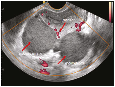 Figura 1.
 Formações císticas retrouterinas de aspecto flocoso fino, sem vascularização interna no Doppler colorido.
A presença de endometriomas ovarianos alerta para minuciosa avaliação dos locais mais frequentes de endometriose profunda, uma vez que são marcadores de endometriose profunda de maior gravidade
(7). A Figura 2 demonstra um endometrioma ovariano. Segundo Guerriero et al.
(9), as lesões podem envolver todo o ovário, permitindo a identificação apenas de pequenas áreas focais periféricas, habitualmente em forma de meia-lua em crescente, que correspondem ao parênquima ovariano residual.
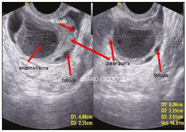 Figura 2.
 Endometrioma no interior do ovário esquerdo. Pode-se observar o parênquima ovariano com folículos na periferia. As setas indicam os folículos ovarianos, o endometrioma e o parênquima.
A presença de nódulos ou lesão hipoecoica irregular, localizados na parede e que envolvem a muscular própria do reto e/ou cólon sigmoide, pode ser considerada como endometriose profunda com comprometimento intestinal
(12). No entanto, para reconhecer, na ultrassonografia, os aspectos da endometriose intestinal, é necessário reconhecer o aspecto ecográfico normal das paredes intestinais (Figura 3).
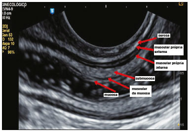 Figura 3.
 Imagem sagital de USTV do reto normal obtida após a preparação do intestino mostra da camada externa para a camada interna.
Gonçalves et al.
(10) consideram haver endometriose intestinal quando a lesão atinge a camada muscular própria. O critério utilizado para predizer esse acometimento foi a presença de nódulo ou espessamento irregular hipoecoico nessa camada da alça, independentemente de a camada hiperecoica, que separa as camadas musculares próprias interna e externa, apresentar solução de continuidade.
As Figuras 4 e 5 ilustram o aspecto característico da endometriose intestinal, representado por uma lesão irregular hipoecogênica.
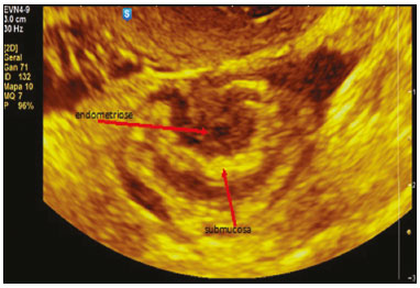 Figura 4.
 Aspecto ecográfico típico de endometriose intestinal (imagem hipoecogênica) acometendo a muscular própria e preservando a camada submucosa.
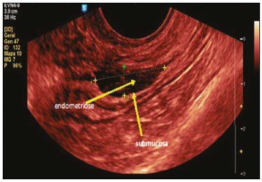 Figura 5.
 Corte sagital demonstrando imagem hipoecogênica infiltrando a serosa e a muscular própria. A submucosa, hiperecogênica, está intacta.
O aspecto ecográfico da endometriose profunda é de espessamento hipoecogênico ou presença de nódulo ou massa com contornos regulares ou irregulares localizados na região retrocervical ou recesso retouterino
(12).
As Figuras 6 e 7 representam o aspecto normal do peritônio uterino posterior (linha hiperecogênica contínua) nos cortes longitudinal e transversal.
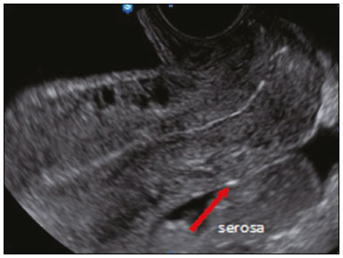 Figura 6.
 Peritônio posterior do útero, corte longitudinal.
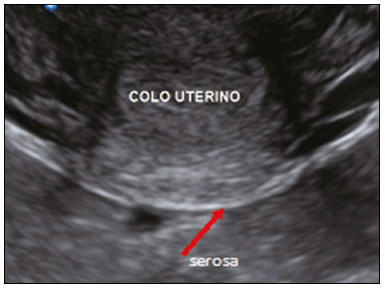 Figura 7.
 Peritônio posterior do útero, corte transversal.
Na Figura 8 é possível constatar que a serosa é interrompida pela lesão, além de infiltrar o colo uterino. Essas situações são frequentemente relatadas em casos de lesões de endometriose profunda encontradas na porção retrouterina, na região retrocervical e no recesso retouterino, infiltrando a parede do fórnice vaginal
(7).
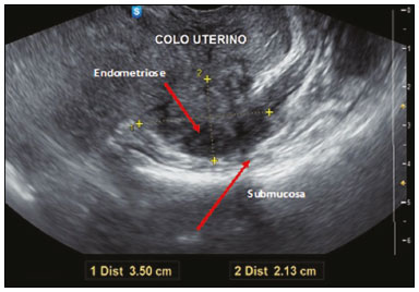 Figura 8.
 Nódulo hipoecogênico sólido, irregular, infiltrando o colo uterino e a muscular própria.
A endometriose ureteral é uma doença rara, mas merece citação pelos sintomas inespecíficos, podendo evoluir silenciosamente para insuficiência renal. É por isso que a avaliação do trato urinário é sugerida quando há suspeita de endometriose profunda infiltrativa e, mais particularmente, se o septo retovaginal é afetado por nódulos maiores que 3 cm
(13).
As lesões retrocervicais são hipoecogênicas infiltrativas e interrompem a linha ecogênica normal que representa o peritônio uterino posterior (Figuras 9 e 10).
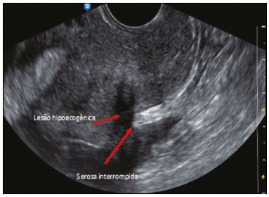 Figura 9.
 Lesão hipoecogênica irregular interrompendo a linha hiperecogênica que representa o peritônio uterino posterior na região do colo uterino.
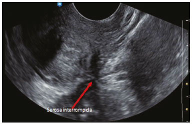 Figura 10.
 Lesão hipoecogênica irregular retrocervical representando lesão endometriótica.
Para endometriose de ligamento redondo, o diagnóstico diferencial principal é o leiomioma subseroso. Na Figura 11 observam-se pequena lesão hipoecogênica no ligamento redondo, que foi descrita como endometrioma porque a paciente apresentava outras lesões de endometriose, inclusive intestinal, e endometrioma à direita. Conforme demonstrado em nosso estudo, lesões como essas são descritas como endometrioma. Especificamente neste caso, a paciente apresentava lesões de endometriose em outros sítios anatômicos.
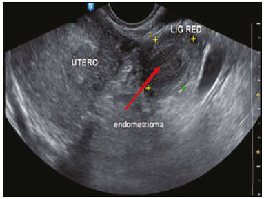 Figura 11.
 Lesão hipoecogênica no ligamento redondo.
O endometrioma ovariano é um marcador para endometriose pélvica e raramente ocorre isoladamente. Assim, ressalta-se que, num contexto clínico em que existe um endometrioma ovariano, a USTV deve investigar a extensão da doença para verificar outras lesões endometrióticas, a fim de escolher o tratamento mais apropriado para tratar a dor e a infertilidade da paciente, não só considerando a presença da lesão ovariana
(14).
Endometrioma na bexigaA USTV é uma técnica precisa para detectar nódulos endometrióticos na parede da bexiga em pacientes com sintomas urinários. Descrevemos um caso caracterizado por lesão nodular hipoecogênica localizada entre a parede uterina anterior do útero e a bexiga (Figuras 12 e 13). Esta paciente engravidou logo após o exame e teve este achado confirmado durante a cesariana.
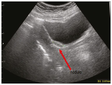 Figura 12.
 Nódulo de endometriose no espaço vesicouterino abaulando a parede vesical, facilmente visualizado pela via transabdominal.
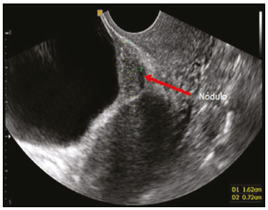 Figura 13.
 Pela via transvaginal, a repleção da bexiga facilita a visualização de nódulo hipoecogênico entre a bexiga e a parede anterior do útero.
A adenomiose define-se pela presença de glândulas endometriais e estroma ao nível da camada muscular uterina. A presença ectópica desse tecido induz hipertrofia e hiperplasia do miométrio envolvente, resultando em aumento do volume uterino. Na maioria dos casos, o tecido endometrial ectópico encontra-se no miométrio em relação com o endométrio eutópico. Porém, pode ocorrer tecido endometrial numa área distante relativamente ao endométrio eutópico, definindo-se, assim, adenomiose subserosa
(15), que pode, raramente, se revelar por um hemoperitônio devido a rotura de quisto
(16).
A Figura 14 mostra um caso característico de adenomiose, formado por um miométrio heterogêneo, sombras em “leque” ou em “raios” e assimetria entre as paredes do útero. Van den Bosch et al.
(17) descrevem a adenomiose como a transformação do endométrio em miométrio, porém, mal definida, com paredes assimétricas e nódulo vascularizado translesional.
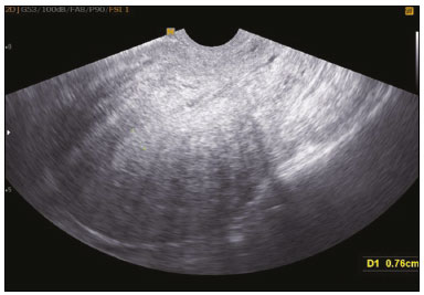 Figura 14.
 Adenomiose, demonstrada pelas sombras “em leque” ou “em raios”.
Os achados ecográficos descritos demonstram a utilidade da ultrassonografia para o diagnóstico da endometriose. Este estudo corrobora outros trabalhos recentes, que indicam a USTV como exame de escolha para ser realizado em mulheres com sintomas suspeitos de endometriose, em virtude da sua simplicidade, boa tolerância e acurácia. Destaca-se, ainda, que o clínico deve ficar atento aos sintomas sugestivos da doença, e os ultrassonografistas realizarem treinamento específico.
REFERÊNCIAS1. Boaventura CS, Rodrigues DP, Silva OAC, et al. Evaluation of the indications for performing magnetic resonance imaging of the female pelvis at a referral center for cancer, according to the American College of Radiology criteria. Radiol Bras. 2017;50:1–6.
2. Alves I, Cunha TM. Clinical importance of second-opinion interpretations by radiologists specializing in gynecologic oncology at a tertiary cancer center: magnetic resonance imaging for endometrial cancer staging. Radiol Bras. 2018;51:26–31.
3. Duarte AL, Dias JL, Cunha TM. Pitfalls of diffusion-weighted imaging of the female pelvis. Radiol Bras. 2018;51:37–44.
4. Godoy LL, Torres US, D’Ippolito G. Subinvolution of the placental site associated with focal retained products of conception and placenta accreta mimicking uterine arteriovenous malformation on CT and MRI: a lesson to be learned. Radiol Bras. 2018;51:135–6.
5. Rodrigues PSC, Silva TSAM, Souza MMT. Endometriose: importância do diagnóstico precoce e atuação da enfermagem para o desfecho do tratamento. Revista Pró-UniverSUS. 2015;6:13–6.
6. Dancet EAF, D’Hooghe TM, Sermeus W, et al. Patients from across Europe have similar views on patient-centred care: an international multilingual qualitative study in infertility care. Hum Reprod. 2012; 27:1702–11.
7. Arruda MS, Camargo MMA, Camargo Jr HSA, et al. Endometriose profunda: aspectos ecográficos. Femina. 2010;38:367–72.
8. Noventa M, Saccardi C, Litta P, et al. Ultrasound techniques in the diagnosis of deep pelvic endometriosis: algorithm based on a systematic review and meta-analysis. Fertil Steril. 2015;104:366–83.
9. Guerriero S, Condous G, van den Bosch T, et al. Systematic approach to sonographic evaluation of the pelvis in women with suspected endometriosis, including terms, definitions and measurements: a consensus opinion from the International Deep Endometriosis Analysis (IDEA) group. Ultrasound Obstet Gynecol. 2016;48:318–32.
10. Gonçalves MO, Dias JA Jr, Podgaec S, et al. Transvaginal ultrasound for diagnosis of deeply infiltrating endometriosis. Int J Gynaecol Obstet. 2009;104:156–60.
11. Pires CR, Zanforlin Filho SM, Lombardi A, et al. Endometriose. In: Pastore A. Ultrassonografia em ginecologia e obstetrícia. Rio de Janeiro, RJ: Revinter; 2006. p. 701–7.
12. Chamié LP, Blasbalg R, Pereira RM, et al. Findings of pelvic endometriosis at transvaginal US, MR imaging, and laparoscopy. Radiographics. 2011;31:E77–100.
13. Donnez J, Nisolle M, Squifflet J. Ureteral endometriosis: a complication of rectovaginal endometriotic (adenomyotic) nodules. Fertil Steril. 2002;77:32–7.
14. Exacoustos C, Pizzo A, Morosetti G, et al. EP27.12: Endometrioma – the tip of a pelvic disease: TVS findings associated with an ovarian endometriosis. Ultrasound Obstet Gynecol. 2016;48(Special Issue):270–393.
15. Sakamoto A. Subserosal adenomyosis: a possible variant of pelvic endometriosis. Am J Obstet Gynecol. 1991;165:198–201.
16. Afonso MC, Castro C, Osório F, et al. Adenomiose: uma apresentação atípica. Acta Obstet Ginecol Port. 2014;8:297–9.
17. Van den Bosch T, Dueholm M, Leone FP, et al. Terms, definitions and measurements to describe sonographic features of myometrium and uterine masses: a consensus opinion from the Morphological Uterus Sonographic Assessment (MUSA) group. Ultrasound Obstet Gynecol. 2015;46:284–98.
1. Promed Clínica Médica, Ijuí, RS, Brasil
2. Secretaria Municipal de Saúde de Ijuí, Ijuí, RS, Brasil
3. Programa de Pós-Graduação em Atenção à Saúde – Universidade de Cruz Alta/Universidade Regional do Noroeste do Estado do Rio Grande do Sul, Cruz Alta/Ijuí, RS, Brasil
a.
https://orcid.org/0000-0001-9558-9441b.
https://orcid.org/0000-0002-4033-3980c.
https://orcid.org/0000-0003-4678-5512d.
https://orcid.org/0000-0003-3631-0847Correspondência:Dr. Jorge Gilmar Amaral de Oliveira
Promed Clínica Médica
Avenida David José Martins, 291
Ijuí, RS, Brasil, 98700-000
E-mail:
gynosom@terra.com.brRecebido para publicação em 7/2/2018
Aceito, após revisão, em 24/4/2018
Data de publicação: 29/08/2019

 |
|


