Sr. Editor,
Paciente feminina, 51 anos de idade, com quadro clínico de cefaleia diária de início insidioso na região frontotemporal esquerda há dois anos, de leve intensidade. Há um ano apresentou o primeiro episódio de crise epiléptica, com característica tônico-clônica generalizada de início focal (parestesias no membro inferior direito), sem fator desencadeante aparente. Faz uso de carbamazepina. Foi admitida pelo departamento de neurologia para investigação de síndrome epiléptica em contexto de síndrome de cefaleia. Apresentava estudo liquórico com marcante hiperproteinorraquia e síntese intratecal de imunogloblulina (IgG) sugerindo componente inflamatório. Foi realizada ressonância magnética (Figura 1).
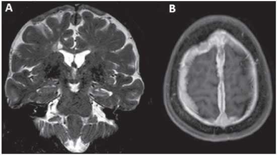
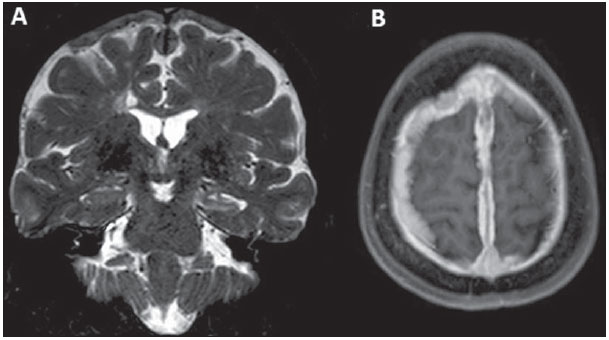
Figura 1. A: Ressonância magnética, coronal T2, mostrando espessamento paquimeníngeo difuso, mais proeminente da alta convexidade, que se estende à foice interhemisférica bilateralmente, com predomínio de hipossinal e associado a redução volumétrica e hipersinal do hipocampo esquerdo (esclerose mesial). B: Axial T1 após administração venosa de contraste paramagnético mostrando realce paquimeníngeo difuso e homogêneo.
A sarcoidose é uma doença multissistêmica, de origem indeterminada, caracterizada por inflamação granulomatosa não caseosa
(1). Há predisposição genética, com ativação dos receptores de linfócitos T por algum antígeno desconhecido. Apresenta predileção pelo sistema respiratório
(1). Nos pulmões, os granulomas são observados no compartimento intersticial e apresentam distribuição perilinfática ao longo das bainhas peribroncovasculares, septos interlobulares e superfície pleural
(1).
A ocorrência da sarcoidose no sistema nervoso central está estimada em torno de 5-15%. É raro o paciente apresentar manifestação neurológica exclusivamente, como no caso aqui apresentado, sendo mais comuns os casos de neurossarcoidose quando há doença disseminada
(2).
As manifestações clínicas da neurossarcoidose são pleomórficas. Comprometimento de nervos cranianos, alterações visuais, cefaleia, fraqueza, paresia, parestesia, alterações psiquiátricas e sinais de irritação meníngea podem ser observados. Sintomas de diabetes insipidus também podem ocorrer, embora sejam mais raros, como sede e poliúria, em razão de acometimento do hipotálamo e hipófise. Quando há acometimento da medula, manifestam-se intensa fraqueza em membros inferiores e outros sinais não específicos de mielopatia
(3).
Embora a neurossarcoidose possa aparecer em todas as regiões do sistema nervoso, é mais comum que ocorra na base do crânio, hipotálamo-hipófise e quiasma óptico
(4). Um dos achados mais comuns na ressonância magnética são as lesões intraparenquimatosas com hipersinal nas sequências em TR longo (T2 e FLAIR), sendo geralmente multifocais periventriculares, subcorticais ou na substância branca profunda. Tais achados são indistinguíveis de vasculites ou de doenças de substrato desmielinizante. Lesões intraparenquimatosas geralmente estão próximas de áreas com acometimento leptomeníngeo (com realce pelo contraste paramagnético) e podem ser únicas ou múltiplas, e envolver também nervos cranianos
(4).
Espessamento paquimeníngeo difuso, com hipossinal em T2, isossinal em T1 e realce pelo meio de contraste, pode também, como no nosso caso, ser observado. Assim, diagnósticos diferenciais, como neurotuberculose, linfoma dural, meningioma em placa, depósitos de IgG4, pseudotumor, metástase de adenocarcinoma, granulomatose de Wegener, paquimeningite hipertrófica idiopática, podem ser considerados, sendo a biópsia necessária para definir a etiologia. É raro o acometimento simultâneo dural e leptomeníngeo
(4). No caso aqui apresentado, os achados de anatomia patológica mostraram granulomas típicos não caseificados na paquimeninge (Figura 2). Outro diagnóstico diferencial a ser considerado é o de hipotensão intracraniana, que apresenta, geralmente, um espessamento paquimeníngeo difuso, porém com hipersinal em T2 (neste caso, havia hipossinal em T2).
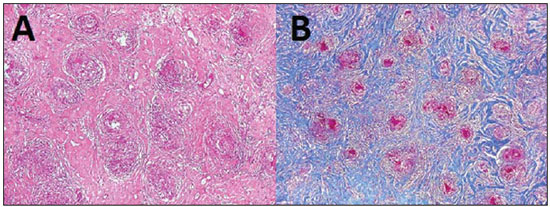
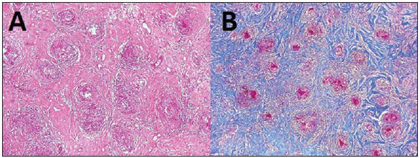
Figura 2. A: Biópsia de área espessada de dura-máter demonstrando numerosos granulomas não caseosos de células epitelioides e gigantes, e infiltrado inflamatório de predomínio linfocitário, em meio a densa fibrose colágena. B: Tricrômico de Masson: destaque dos granulomas contra o denso tecido conjuntivo colágeno.
Ainda não há um consenso sobre o tratamento da neurossarcoidose. Quando o paciente é sintomático, iniciam-se altas doses de corticoide, que podem ser reduzidas ao longo do tratamento e até retiradas
(3).
REFERÊNCIAS1. Melo ASA, Marchiori E, Capone D. Tomographic and pathological findings in pulmonary sarcoidosis. Radiol Bras. 2011;44:220-4.
2. Spencer TS, Campellone JV, Maldonado I, et al. Clinical and magnetic resonance imaging manifestations of neurosarcoidosis. Semin Arthritis Rheum. 2005;34:649-61.
3. Nozaki K, Judson MA. Neurosarcoidosis: clinical manifestations, diagnosis and treatment. Presse Med. 2012;41(6 Pt 2):e331-48.
4. Christoforidis GA, Spickler EM, Recio MV, et al. MR of CNS sarcoidosis: correlation of imaging features to clinical symptoms and response to treatment. AJNR Am J Neuroradiol. 1999;20:655-69.
Universidade Estadual de Campinas (Unicamp), Campinas, SP, Brasil
Endereço para correspondência:
Dr. Fabiano Reis
Faculdade de Ciências Médicas - Universidade Estadual de Campinas, Departamento de Radiologia
Rua Tessália Vieira de Camargo, 126, Cidade Universitária Zeferino Vaz
Caixa Postal: 6111. Campinas, SP, Brasil, 13083-887
E-mail:
fabianoreis2@gmail.com

 |
|


 Read in English
 PDF Portuguese
 Print
Print
 Send this article by email
 How to cite this article
 Submit a comment
 Mendeley
 Pocket