Radiologia Brasileira - Publicação Científica Oficial do Colégio Brasileiro de Radiologia
AMB - Associação Médica Brasileira CNA - Comissão Nacional de Acreditação
 Vol. 48 nº 1 - Jan. /Feb. of 2015
|
ORIGINAL ARTICLE
|
|
Ambient radiation levels in positron emission tomography/computed tomography (PET/CT) imaging center |
|
|
Autho(rs): Priscila do Carmo Santana1; Paulo Marcio Campos de Oliveira1; Marcelo Mamede2; Mariana de Castro Silveira3; Polyanna Aguiar3; Raphaela Vila Real3; Teógenes Augusto da Silva4 |
|
|
Keywords: Dosimetry; Thermoluminescence dosimetry; PET/CT. |
|
|
Abstract: INTRODUCTION
The adoption of ambient radiation monitoring programs is in general aimed at assessing the radiological conditions in the workplace. The ambient monitoring program will ensure that work conditions are acceptably safe and satisfactory for exposed individuals and that the dose levels established by the regulatory authorities (in Brazil, Comissão Nacional de Energia Nuclear - CNEN), both for free and controlled areas, are not exceeded(1). Ambient dosimetry is an integral part of the ambient monitoring program. Such a dosimetry is necessary to estimate the doses in locations where there might exist exposure to ionizing radiations, both for occupationally exposed individuals and patients, as well as for the public in general. The preoccupation with ambient dosimetry is observed in all radiodiagnosis modalities. Adad et al. have determined the isodose curve in a mammography room and concluded that the utilization of additional shielding at mammography rooms is not necessary, since at distances > 0.50 m, the measurements generated absorbed doses < 0.1 mGy per exposure(2). Vieira et al. (Determination of isodose curves in brachytherapy with linear radiation sources. VI Congressso Brasileiro de Física Médica; 2001 Oct 4-6; Rio de Janeiro, Brazil) have determined isodose curves in brachytherapy for linear radiation sources, while Goulart et al. (Determination of isoexposure curves from a digital fluoroscopy apparatus at a hemodynamics room. VIII Congresso Brasileiro de Física Médica; 2003 May 13-16; Porto Alegre, Brazil) have determined isodose curves in a hemodynamics room, and Andrade et al. (Determination of isoexposure curves in patients submitted to iodine therapy. VIII Congresso Brasileiro de Física Médica; 2003 May 13-16; Porto Alegre, Brazil) have estimated isodose curves for a digital fluoroscopy apparatus and also in the room where patients received iodine therapy doses in nuclear medicine. Avila et al. have determined the ambient dose at a nuclear medicine service with TLD-100 and TLD-900 detectors. In the gamma chamber, the rate of ambient dose equivalent was approximately 0.05 µSv/h. In the other monitored locations, with the two types of detectors, the ambient dose equivalent values with TLD-900 detector were 25%-45% higher than the values observed with the utilization of the TLD-100 detector. Such a result was attributed to the production of low-energy scattered radiation that results in greater response from the TLD-900 detector and, therefore, the values found with the TLD-100 detector were considered to be more reliable(3). PET/CT apparatuses are dedicated to the study of positron emitters (for example: 18F, 11C, 15O). At PET/CT-based nuclear medicine centers, the most commonly found radioisotope is fluorine-18-labeled fluorodeoxyglucose (18F-FDG), whose energy released after interaction with the medium is 511 keV, with a half-life of approximately 109 minutes. CT, which was incorporated into this technology, is basically utilized for attenuation correction in organs surrounding the region of interest and to assist in the accurate anatomical localization of the molecular changes identified by PET. Imaging diagnosis centers specialized in the PET/CT technique must comply with annual dose restriction values (5 mSv for controlled and supervised areas, and 0.5 mSv for free areas) established by the standard CNEN NN 3.01 - "Directives for radiological protection"(1). With a view to assuring radiological protection for workers and general public individuals, dosimetry was carried out at Centro de Imagem Molecular (CImol) (Molecular Imaging Center), a building located beside the Faculdade de Medicina da Universidade Federal de Minas Gerais complex. The dosimetry comprised the entire area occupied by CImol, as well as adjacent rooms. According to Ordinance 453/98 from Agência Nacional de Vigilância Sanitária (Brazilian Health Surveillance Agency), "Directives for radiological protection in dental and medical radiodiagnosis" the areas should be classified into free areas and controlled areas and the dose restriction levels should be 0.5 mSv/year and 5.0 mSv/year, respectively(4). On the other hand, the standard CNEN NN 3.01 - "Radiological Protection Basic Directives" - establishes that the services must assess the levels of dose restriction compatible with their activities as a limiting condition for the process of radiological protection optimization(1). In the publication AAPM-108 - "PET and PET/CT shielding requirements" -, the American Association of Physicists in Medicine recommends the utilization of the 5.0 mSv/year value for the purpose of shielding calculation, in order to optimize the radiation level to which occupationally exposed individuals are subjected(5). The present study was aimed at demonstrating that the radiation levels in areas occupied by professionals operating PET/CT equipment, workers in adjacent areas and general public individuals are compatible with the thresholds for external exposure established by more restrictive standards such as Ordinance 453/98, provided appropriate radioprotection measures are adopted. MATERIALS AND METHODS CIMol is a PET/CT imaging center that complies with the recommended radioprotection requirements, periodically performing ambient monitoring with thermoluminescent detectors in order to assure that the radiation levels are within recommended limits. As a part of Instituto Nacional de Ciência e Tecnologia em Medicina Molecular, CIMol provides nuclear medicine services, occupying an area of approximately 320 m2, is equipped with a GE Discovery 690 PET/CT apparatus with Lyso detectors technology and a 64-channel (multislice) CT apparatus. The PET/CT installation is located on the first floor, comprising two toilets for workers, a small coffee break room, meeting room and reporting room, all of these being considered free areas. The controlled area comprises a source handling room, a control room, the PET/CT apparatus room, machinery room, nursing station, three exclusive toilets for patients, four activation boxes and a waste area. The access to the controlled area is only possible by means of digital identification. The second floor comprises a research laboratory and offices where workers spend about eight hours per day. The entire CIMol facility and surrounding areas were monitored with the utilization of TLD-100H magnesium-, copper- and phosphor-doped (LiF:Mg,Cu,P) lithium fluoride thermoluminescent dosimeters calibrated for the equivalent dose range for photons (Hx), whose detection threshold is 5 × 10⁻³ mSv. The dosimeters calibration was carried out at the Dosimeter Calibration Laboratory of Centro de Desenvolvimento da Tecnologia Nuclear, a research institute from CNEN. The decision to utilize the TLD-100H detectors was based on some advantageous features, among them the high sensitivity to gamma radiation, 40 times greater than that of other thermoluminescent detectors(6). The ambient dosimetry measurements were obtained by exposing the TLD-100H dosimeters over a period of 32 consecutive days at the points indicated on the CIMol facility floor plan as shown on Figures 1 and 2. Each point was selected on the basis of its occupation by workers and general public individuals, and the radiation levels were evaluated with three thermoluminescent detectors in order to assure greater metrological reliability. The detectors were positioned in the interior of the premises, protected by a 1.4 m tall support reproducing the most exposed region of the chest of an average size adult. 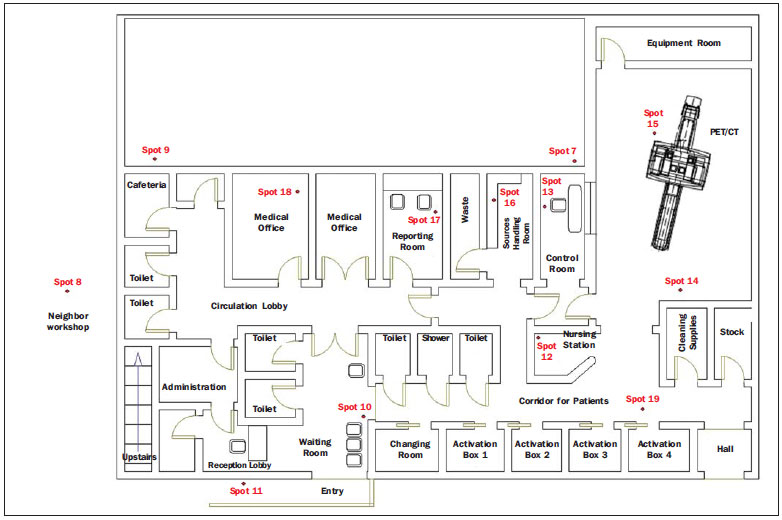 Figure 1. Sketch of the first floor of Centro de Tecnologia em Imagem Molecular (CIMol). 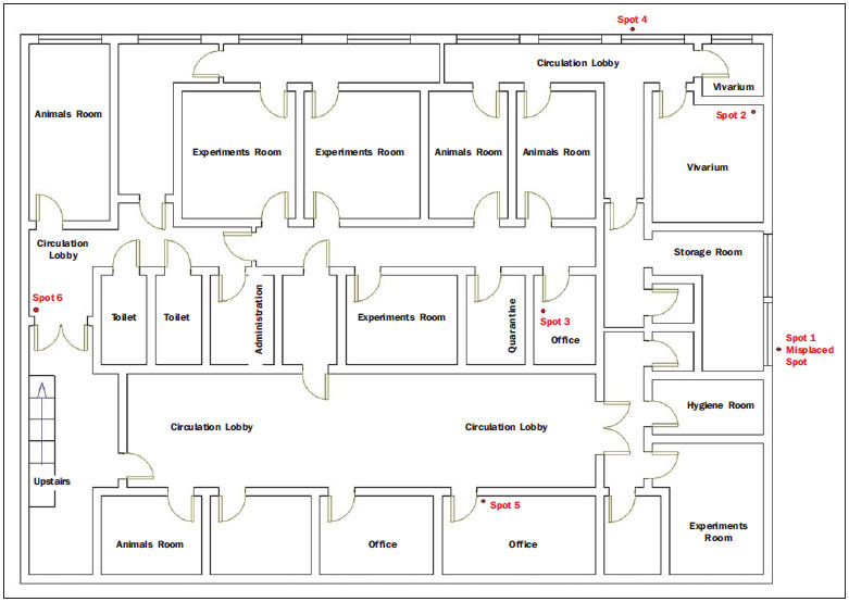 Figure 2. Sketch of the second floor of Centro de Tecnologia em Imagem Molecular (CIMol). The calculation of the annual dose estimates from the results in terms of dose equivalent for photons (Hx) evaluated in the 32 day interval, was made according to equation, as follows: 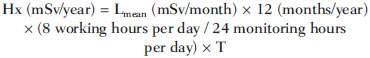 where: Hx is the dose equivalent for photons in mSv/year; Lmean is the mean value of the readings in mSv; T is the area occupation factor, based on the mean stay time of a person in that location. The measurement results were followed by the respective standard expanded uncertainty values and the main considered contribution sources were the measurements reproducibility (type A uncertainty) and the intrinsic dosimeters' features, such as energetic and angular dependence, besides the imported calibration uncertainties. All calculations were performed on the basis of the Brazilian recommendations(7). An average number of 12 PET/CT studies per week were performed, a number that may vary slightly on a monthly basis, but not significantly over a one-year period. It is important to highlight that, after the radiopharmaceutical injection, the patient remains for approximately 50 minutes in an activation box, being then swiftly moved through an internal circulation corridor that leads to a toilet and to the PET/CT apparatus. The patient remains in the toilet for approximately 2 minutes and is then moved to the examination room, where he or she stays for a mean period of 30 minutes for acquisition of the PET images. For the acquisition of the CT images, the parameters of 120 kV, with modulated current (between 10 and 150 mA) are utilized according to patient size and scan type, with slice thickness varying according to the evaluated region (0.625 to 20 mm) and slice time of 0.5 to 1.0 s. RESULTS After the measurements were taken, the ambient dose was calculated, and Table 1 presents the estimates of external annual dose (Hx) for each monitored point with the respective measurement uncertainty value. 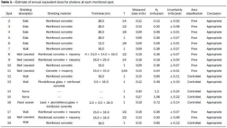 DISCUSSION At the spots 2, 4, 6, 9 and 11, the dose estimates indicated values of up to 44% of the restriction level of annual dose for free areas (0,5 mSv/ano). The spots 3, 5, 7, 8, 10, 17 and 18 presented annual dose estimates reaching values between 72% and 87% of the restriction level of annual dose for free areas. Such results demonstrated that the shielding of such areas is appropriate for the current demand of the center. It is important to highlight that the CNEN standards (CNEN-NN-3.01 and CNEN-NN-3.05) do not define ambient dose restriction values for nuclear medicine centers, thus the dose restriction levels established by the Ministry of Health (Ordinance 453/98) for medical radiodiagnosis services were utilized. Such levels were considered as photons are utilized at a lower magnitude energy range, and therefore, the levels are more restrictive, thus favoring radiological safety. At spots 12 to 16, and spot 19, all of them in controlled areas, the dose estimates presented values of up to 29% of the annual dose restriction level for controlled areas (5.0 mSv/year), indicating that such spots are appropriate in terms of radiation level, and that the shielding, as applicable, are appropriate for the current demand of the center. In all compliance evaluations with respect to dose restriction, the results for dose equivalent for photons in one year corresponded to the sum of the mean readings from the dosimeters with their respective uncertainty values. It is important to highlight that the results were overestimated for both the Hx evaluation for the evaluation of uncertainties, according to the radiological protection principles. Based on the present results, it is possible to conclude that there is no need to make any changes in the physical structure of the center, since in none of the spots the thresholds established by the current standards were exceeded. Also, based on the time of equipment utilization and image acquisition parameters, it is possible to conclude that the number of exams carried out at CIMol might be increased by 10%. CONCLUSION The results from the evaluation of radiation levels for both the internal and external areas of CIMol demonstrate that the dose estimates for the areas are appropriate in relation to dose restrictions established by the Ministry of Health, as the CNEN regulations do not establish such values for nuclear medicine centers. The values for all the spots in the center were found to be satisfactory, demonstrating that the whole shielding system is appropriate for both the free and controlled areas. Also, it is possible to conclude that the number os exams performed at CIMol might be increased by 10%. And, considering that the center operates with unsealed radioactive sources, one should stress the importance of strict compliance with radioprotection principles. REFERENCES 1. Brasil. Ministério da Ciência, Tecnologia e Inovação. Comissão Nacional de Energia Nuclear. Diretrizes básicas de proteção radiológica. CNEN-NN-3.01, de 1º de setembro de 2011. 2. Adad MCBT, Hoff G, Streck EE, et al. Curvas de isodose no ar em uma sala de mamografia. Radiol Bras. 2008;41:255-8. 3. Avila O, Torres-Ulloa CL, Medina LA, et al. TL measurement of ambient dose at a nuclear medicine department. Radiation Measurements. 2011;46:1843-6. 4. Brasil. Ministério da Saúde. Secretaria de Vigilância Sanitária. Diretrizes de proteção radiológica em radiodiagnóstico médico e odontológico. Portaria nº 453, de 1º de junho de 1998. 5. Madsen MT, Anderson JA, Halama JR, et al. AAPM Task Group 108: PET and PET-CT shielding requirements. Med Phys. 2006;33:4-15. 6. Oliveira ML, Maia AF, Nascimento NCES, et al. Influência da dependência energética de dosímetros termoluminescentes na medida da dose na entrada da pele em procedimentos radiográficos. Radiol Bras. 2010;43:113-8. 7. Instituto Nacional de Metrologia, Qualidade e Tecnologia. Avaliação de dados de medição: guia para a expressão de incerteza de medição - GUM 2008. 1ª ed. Duque de Caxias, RJ: Inmetro/Cicma/Sepin; 2012. 1. PhDs, Associate Professors, Universidade Federal de Minas Gerais (UFMG), Belo Horizonte, MG, Brazil 2. PhD, Full Professor, Universidade Federal de Minas Gerais (UFMG), Belo Horizonte, MG, Brazil 3. Graduate Students, Course of Technology in Radiology, Universidade Federal de Minas Gerais (UFMG), Belo Horizonte, MG, Brazil 4. PhD, Titular Researcher at Centro de Desenvolvimento da Tecnologia Nuclear - Comissão Nacional de Energia Nuclear (CDTN/CNEN), Belo Horizonte, MG, Brazil Mailing Address: Dra. Priscila do Carmo Santana Departamento de Anatomia e Imagem - UFMG Avenida Professor Alfredo Balena, 190, Santa Efigênia Belo Horizonte, MG, Brazil, 30130-100 E-mail: pridili@gmail.com Received June 11, 2013. Accepted after revision May 8, 2014. Study developed in the Department of Anatomy and Imaging, Universidade Federal de Minas Gerais (UFMG), Belo Horizonte, MG, Brazil. |
|
Av. Paulista, 37 - 7° andar - Conj. 71 - CEP 01311-902 - São Paulo - SP - Brazil - Phone: (11) 3372-4544 - Fax: (11) 3372-4554