Radiologia Brasileira - Publicação Científica Oficial do Colégio Brasileiro de Radiologia
AMB - Associação Médica Brasileira CNA - Comissão Nacional de Acreditação
 Vol. 47 nº 4 - July / Aug. of 2014
|
ORIGINAL ARTICLE
|
|
Optimization of a protocol for myocardial perfusion scintigraphy by using an anthropomorphic phantom |
|
|
Autho(rs): Susie Medeiros Oliveira Ramos1; Adriana Pereira Glavam2; Tadeu Takao Almodovar Kubo3; Lidia Vasconcellos de Sá4 |
|
|
Keywords: Myocardial perfusion imaging; Optimization; Anthropomorphic phantom. |
|
|
Abstract: INTRODUCTION
Coronary arterial disease (CAD) is the main cause of death among men and women in the world, according to the World Health Organization(1). Estimates indicate that approximately one individual dies every 40 seconds on account of cardiovascular diseases(2). The greater prevalence of CAD in male individuals caused this disease to be underdiagnosed and undertreated in women, in spite of being an important morbimortality factor in this population. This is due to the fact that the greatest part of the studies in the literature approaching diagnosis, treatment and prognosis of CAD has contemplated a relatively small number of women. Thus, the obtained results in such studies cannot always be extrapolated to this population. The diagnosis of CAD in women is always a challenge. The clinical presentation is late (approximately 10 to 15 years after the men's) and the symptoms are usually atypical. For being more elderly, women present with a greater number of comorbidities and worse prognosis. It is important to remind that the prevalence of CAD after the 7th decade of life is similar in men and women, and once it is diagnosed, the prognosis is worse in women. In developed countries, more than 50% of the women die on account of cardiovascular diseases, and sudden deaths are responsible for 35% of the mortality(3). Myocardial perfusion scintigraphy (MPS) is a noninvasive imaging method with high diagnostic and prognostic value, and it is widely validated in clinical practice both for men and women(46). Such nuclear medicine imaging method relies on the utilization of a gamma camera and a radiopharmaceutical. Currently, 99mTc-sestamibi is the most used radiopharmaceutical in this procedure. The two-day protocol includes recommended activities of 888 to 1,332 MBq, according to the American Society of Nuclear Cardiology(7), and 600 to 900 MBq, as stated by the European Council on Nuclear Cardiology (ECNC), that also recommends a time of 25 seconds per acquisition as ideal for cardiac SPECT(8). The present study was aimed to the optimization of 99mTc-sestamibi MPS by varying the activities and acquisition times, and verifying the influence of such parameters on the image quality in a cardiac anthropomorphic phantom. MATERIALS AND METHODS Anthropomorphic phantom A Data Spectrum ECT/TOR/P model anthropomorphic phantom was utilized, simulating the upper part of a medium to large-sized individual's chest. Such a phantom comprises heart simulators (with the possibility of attaching lesions of different sizes), lungs, liver, and dorsal spine (Figure 1). 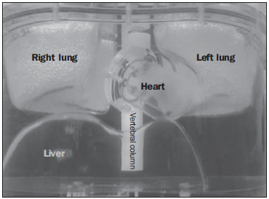 Figure 1. Anterior view of the ECT/TOR/P model anthropomorphic phantom, Data Spectrum Corporation. Equipment and imaging protocol The nuclear medicine imaging system was a GE Healthcare Ventri model gamma camera with SPECT (single photon emission tomography) comprising a fixed 90° dual-head system and an image processing software, Xeleris2 (release 2.151). Each detector has a 370 × 190 mm rectangular field of view (FOV) of a 9.5 mm-thick NaI(Tl) crystal. The two-day protocol included the following characteristics:
Different activities were utilized not only to reduce the doses received by patients, respecting the international ALARA (as low as reasonably achievable) concept, but also to meet possible restrictions in the radiopharmaceuticals supply, as it recently occurred during the so-called "generator crisis" (2010/2011). Image acquisitions were also performed with different projection times, in order to analyze whether the usual acquisition time could be optimized, both for the patient's comfort as well as for the reduction of motion artifacts which may lead to scan repetition. Simulated variations
Image processing and reconstruction The images were iteratively reconstructed with the aid of the software Evolution for Cardiac from GE Healthcare, a recently introduced algorithm for cardiac images reconstruction. Such algorithm incorporates RR (resolution recovery) and MAP (maximum a posteriori) type noise regularization, allowing the SPECT images to be acquired in half the time compared with standard OSEM (ordered subset expectation maximization) algorithm, being known as HT (half time acquisition)(9). Preparation of the anthropomorphic phantom Measurements were performed with and without insertion of heart lesions, in order to simulate healthy individuals and the ones with CAD. For the studies that simulated hypo-uptake, a 1.0 cm-thick solid-type lesion with 2 cm in length was inserted in the lower basal region of the cardiac phantom. The radioactivity concentration in the cardiac phantom followed the pharmaceutical biodistribution(10,11). The details of the activity in each organ are individually presented on Table 1. The concentration ratio (MBq/ml) between heart:liver:body was 12:8:1. According to the literature, the concentration ratio (MBq/ml) myocardium/liver is 1.3 ± 0.1(1012). 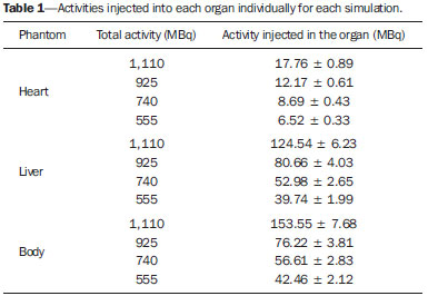 Image evaluation method Qualitative and semi-quantitative analyses: scores on medical evaluation The clinical evaluation was blindly performed by an experienced nuclear cardiologist, i.e., without any knowledge on the parameters utilized in the images acquisition. The scores considered three criteria, as follows:
The semi-quantitative evaluation is aimed at standardizing the segmental analysis of the left ventricle (LV) and the lower subjectivity in the interpretation. A system of scores for the 17 segments of the LV which considers three sections in the smaller axis (apical, median and basal) and a section on the long vertical axis, is utilized as shown on Figure 2. 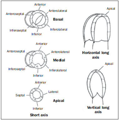 Figure 2. Image of left ventricle with recommended terminology of the 17 segments for tomographic images of the heart. Each one of the 17 segments is scored according to the radiopharmaceutical uptake, as follows: 0 (zero) = normal; 1 (one) = slight reduction in the radiopharmaceutical uptake; 2 (two) = moderate reduction in the radiopharmaceutical uptake; 3 (three) = significant reduction in radiopharmaceutical uptake; 4 (four) = absence of radiopharmaceutical uptake. The sum of the values attributed to each representative segment of the stress phase is called SSS. The values between 0 and 4 are considered as normal or equivocal (possibility of attenuation artifact) and SSS values > 4 are considered as altered. Semi-quantitative analysis with software The software analysis was performed with the aid of the software ImageJ. Short and long vertical axes images were selected for analysis resulting from processing, with 600% zoom. Customized regions of interest (ROI) were delimited for each one of the 17 segments. Such ROI were defined with the assistance of a nuclear cardiologist, being repeated in an identical manner in all heart images. The sum of the pixel count values in each ROI were graphically represented, comparing the phantom with a patients' data bank. Validation of the method In order to validate the data obtained with the phantom, the results were compared with those from a group of female patients with pre-test low probability of CAD, according with the criteria by Diamond et al.(12). A total of 40 studies were used and originated a databank representative of the normal radiotracer uptake on each one of the analyzed 17 LV segments. The mean age of the patients selected as standard databank was 57.3 years (3669 years); mean body mass index of 25.40 (1937); and mean injected activity of 811.78 MBq (699.301,013.80 MBq). Such data were collected from the databank of the clinic where the study was performed. RESULTS Initially, the activities and acquisition times were varied in the phantom without heart lesions. The scores from the evaluations performed by the cardiologist regarding technical image quality, evaluation of perfusion and SSS value (observer-dependent) obtained for activities from 555 to 925 MBq and acquisition times from 7 to 20 seconds per projection can be observed on Table 2. 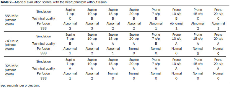 The investigation performed with a lesion in the inferobasal region of the heart was not performed with 555 MBq (15 mCi) due to the results from the first part of the study. Likewise, the images were not acquired in the prone position with more than 7 seconds per projection. The cardiologist evaluation scores for simulation with lesion can be seen on Table 3. 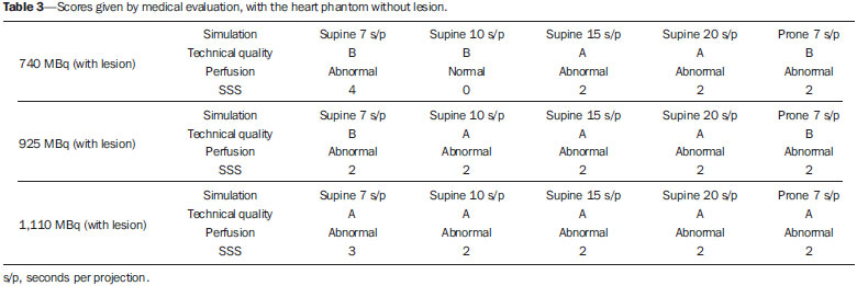 By simulating the usually administered activity of 1,110 MBq, the acquisition time could be reduced to 7 seconds per projection, which is a reduction of 53.34% of the time used in the standard protocol, without changing the possibility of identifying the lesion, as demonstrated on Figure 3. 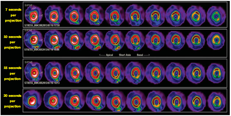 Figure 3. Processed images of the heart phantom with a lesion in the inferobasal segment, with times varying between 7 and 20 seconds per projection, highlighting that the protocol's standard time is 15 seconds. The ROI delineated for quantification at ImageJ can be observed on Figure 4. The counts for each segment were used to compare with the data from patients in the normality databank. 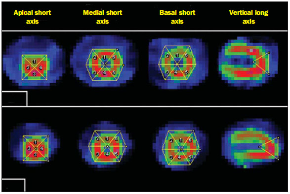 Figure 4. Regions of interest delineated for each one of the 17 segments of the heart by the ImageJ software. A: Anthropomorphic heart phantom. B: Patient's exam. In order to validate the data, the counts for each segment of the heart phantom were compared with those from the normality databank by the ImageJ software. The chart on Figure 5 shows such comparison. 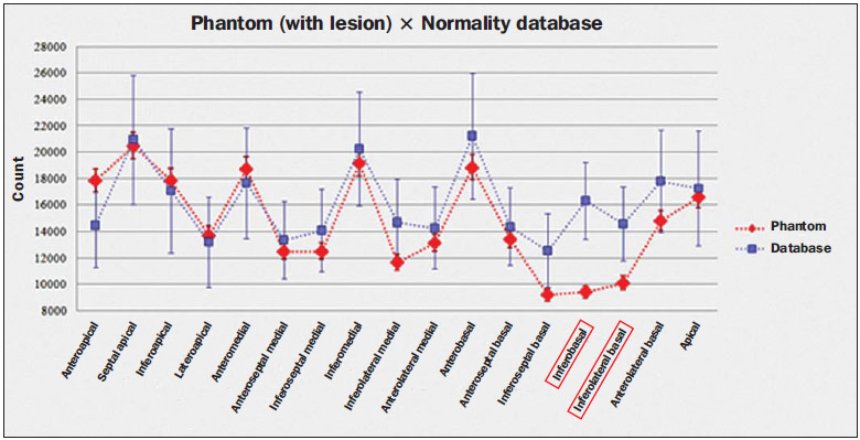 Figure 5. Count values at the 17 segments compared with the databank. The lesion on the heart phantom is observed as well as its influence on neighboring segments. The lesion positioned in the inferobasal region of the phantom was the only count with a value below those of the databank, as this comprised data from healthy patients. The injected activity in the phantom was 1,110 MBq, demonstrating that this is the actual activity administered in the patients. DISCUSSION The results demonstrated that the scan cannot be performed with 555 MBq (15 mCi) of injected activity, as the technical quality of the image is not appropriate for an accurate diagnosis, according to the acquisition protocols adopted in the present study. In the images acquired with the phantom in ventral decubitus (prone), with 740, 925 and 1,110 MBq, no change was observed in the image quality, even for acquisitions with 7 seconds per projection. It was observed that the injected activity could be reduced to 740 MBq (33.34% reduction) if the acquisition time remained the same as the usual time of 15 seconds per projection. For a time shorter than this, the lesion may be masked by the lack of counting statistics. Nevertheless, the activity could be reduced to 925 MBq (16.67% reduction) and 53.34% reduction in acquisition time with no change in image quality and with the lesion still being properly diagnosed. With the usual injected activity of 1,110 MBq, the acquisition time could be reduced to 7 seconds per projection both in the supine and in the prone positions (reduction of 53.34%), without any change in image quality and in the diagnostic accuracy of the method. The chart on Figure 5 shows the count on each one of the 17 heart segments for the normality databank, considering the count on the same segments of the heart phantom. The values observed were within the mean value of the counts considering the standard deviation, which confirms the validity of the proposed phantom usage. CONCLUSION The present study demonstrated that the ECT/TOR/P Data Spectrum anthropomorphic phantom appropriately reproduces patients, within the mean and standard deviation of a randomly selected sample. After validation, activity and time parameters were varied for the two-day protocol study in MPS. The results demonstrated that for the same time currently used for the exam, the injected activity could be reduced in up to 33.34%. If the acquisition time is also reduced (to 53.34% of the usual time), the activity could be reduced up to 16.67% of the usual injected activity, without any change in the image quality and in the diagnosis. The images acquired in ventral decubitus (prone) demonstrated improvement in relation to those acquired only in supine position, complementing the exam. A deeper study of optimization based on positioning is currently underway and should produce more results for the present investigation on the two-day protocol for this type of exam. Such results serve as grounds for the undertaking of clinical studies involving patients for definition of an optimized protocol. REFERENCES 1. World Health Organization, World Heart Federation, World Stroke Organization. Global atlas on cardiovascular disease prevention and control. [acessado em 8 de agosto de 2013]. Disponível em: http://www.who.int/cardiovascular_diseases/publications/atlas_cvd/en/. 2. Go AS, Mozaffarian D, Roger VL, et al. Heart disease and stroke statistics 2013 update: a report from the American Heart Association. [acessado em 2 de outubro de 2013]. Disponível em: http://circ.ahajournals.org/content/127/1/e6.full. 3. Issa AFC, Pantoja MR. Valor prognóstico da cintigrafia de perfusão miocárdica em mulheres comparado com homens com suspeita clínica de doença coronariana. Rev SOCERJ. 2006;19:919. 4. Henzlova MJ, Cerqueira MD, Hansen CL, et al. ASNC imaging guidelines for nuclear cardiology procedures: stress protocols and tracers. [acessado em 3 de junho de 2013]. Disponível em: https://www.asnc.org/imageuploads/imagingguidelinesstressprotocols021109.pdf. 5. Hesse B, Tägil K, Cuocolo A, et al. EANM/ESC procedural guidelines for myocardial perfusion imaging in nuclear cardiology. Eur J Nucl Med Mol Imaging. 2005;32:85597. 6. Ali I, Ruddy TD, Almgrahi A, et al. Half-time SPECT myocardial perfusion imaging with attenuation correction. J Nucl Med. 2009;50:55462. 7. Savi A, Gerundini P, Zoli P, et al. Biodistribution of Tc-99m methoxy-isobutyl-isonitrile (MIBI) in humans. Eur J Nucl Med. 1989;15:597600. 8. Münch G, Neverve J, Matsunari I, et al. Myocardial technetium-99m-tetrofosmin and technetium-99m-sestamibi kinetics in normal subjects and patients with coronary artery disease. J Nucl Med. 1997;38:42832. 9. Zolle I. Technetium-99m pharmaceuticals: preparation and quality control in nuclear medicine. Berlin Heidelberg: Springer; 2007. 10. Bucerius J, Ahmadzadehfar H, Biersack HJ. 99Tc-sestamibi: clinical applications. Berlin Heidelberg: Springer; 2012. 11. Cerqueira MD, Weissman NJ, Dilsizian V, et al. Standardized myocardial segmentation and nomenclature for tomographic imaging of the heart. A statement for healthcare professionals from the Cardiac Imaging Committee of the Council on Clinical Cardiology of the American Heart Association. Ciculation. 2002;105:53942. 12. Diamond GA, Forrester JS. Analysis of probability as an aid in the clinical diagnosis of coronary artery disease. N Engl J Med. 1979;300:13508. 1. Biomedical Researcher, Master in Radioprotection and Dosimetry, Instituto de Radioproteção e Dosimetria / Comissão Nacional de Energia Nuclear (IRD/CNEN), Rio de Janeiro, RJ, Brazil 2. Master in Cardiology, Nuclear Cardiologist, Clínica de Diagnóstico Por Imagem (CDPI/DASA), Rio de Janeiro, RJ, Brazil 3. Master, Medical Physicist specialized in Images Post-processing and Nuclear Medicine, Clínica de Diagnóstico Por Imagem (CDPI/DASA), Hospital Unimed Rio and Phys RAD, Rio de Janeiro, RJ, Brazil 4. PhD, Senior Technologist, Instituto de Radioproteção e Dosimetria / Comissão Nacional de Energia Nuclear (IRD/CNEN), Rio de Janeiro, RJ, Brazil Mailing Address: Susie Medeiros Oliveira Ramos Instituto de Radioproteção e Dosimetria, Departamento de Física Médica Avenida Salvador Allende, s/nº, Jacarepaguá Rio de Janeiro, RJ, Brazil, 22780-160 E-mail: susie@ird.gov.br Received September 29, 2013. Accepted after revision February 10, 2014. Study developed at Instituto de Radioproteção e Dosimetria / Comissão Nacional de Energia Nuclear (IRD/CNEN), Rio de Janeiro, RJ, Brazil. Financial support: Coordenação de Aperfeiçoamento de Pessoal de Nível Superior (Capes) / Comissão Nacional de Energia Nuclear (CNEN) Master's scholarship. |
|
Av. Paulista, 37 - 7° andar - Conj. 71 - CEP 01311-902 - São Paulo - SP - Brazil - Phone: (11) 3372-4544 - Fax: (11) 3372-4554