Radiologia Brasileira - Publicação Científica Oficial do Colégio Brasileiro de Radiologia
AMB - Associação Médica Brasileira CNA - Comissão Nacional de Acreditação
 Vol. 46 nº 6 - Nov. / Dec. of 2013
|
WHICH IS YOUR DIAGNOSIS?
|
|
Which is your diagnosis? |
|
|
Autho(rs): Gláucia Zanetti1; Luiz Felipe Nobre2; Alexandre Dias Mançano3; Marcos Duarte Guimarães4; Bruno Hochhegger5; Arthur Soares Souza Jr.6; Edson Marchiori1 |
|
|
A 59-year-old female patient presented with fever and dry cough. Chest radiography taken at admission to the emergency department showed pulmonary infiltrate. The patient was referred to the hospital to undergo highresolution computed tomography scan (Figure 1).
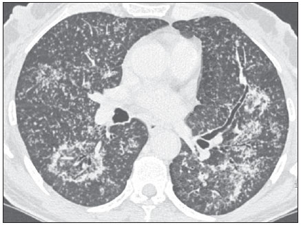 Figure 1. High-resolution computed tomography, lower pulmonary regions section. Image description Figure 1. High-resolution CT image of lower pulmonary regions shows numerous small, bilateral, random nodules, clusters of small nodules in the left lung, and reversed halo sign in the superior segment of the right lower lobe. Note the nodular walls of the lesion and the presence of small nodules within the lesion. Diagnosis: Nodular reversed halo sign caused by pulmonary tuberculosis, confirmed by sputum culture. COMMENTS Reversed halo sign (RHS) is defined as a focal, round area of ground-glass attenuation surrounded by a partial or complete rim of consolidation(1). It was initially described as a relatively specific sign of cryptogenic organizing pneumonia. However, subsequent publications have identified this sign in a wide spectrum of diseases, including infectious and non-infectious conditions(26). Although RHS must be regarded as a non-specific sign that is found in various pulmonary diseases, authors have observed that, in cases of active granulomatous diseases presenting the RHS, the rim of the reversed halo may be nodular in appearance(7). Most of the reported cases of RHS related to a proven granulomatous infection such as tuberculosis(810) or active sarcoidosis(1115) exhibited a nodular rim. Additionally, in general, small nodules are observed in the center of the reversed halo signs. In such cases, histopathological analysis has revealed the presence of granulomas both in the ring and within the reversed halo signs. Recently, a comparative study of 12 cases of RHS in patients with tuberculosis and 10 in patients with COP(8) demonstrated that all patients with tuberculosis presented nodular RHS, and also that small nodules were observed in the ground-glass component of the RHS in 83% of those cases. No patient with COP presented nodular RHS or central nodules. The importance of identifying imaging patterns that could raise the possibility of active tuberculosis has long been recognized as highly relevant for public health and to ensure that infected patients receive the appropriate therapy. Acid-fast bacilli are found in the sputum in only a limited number of patients with active pulmonary tuberculosis. For this reason, antituberculosis treatment is frequently initiated and preventive measures such as patient isolation are taken on the basis of imaging findings suggestive of active tuberculosis even before bacteriological confirmation(16). Well-recognized HRCT findings of postprimary pulmonary tuberculosis include centrilobular or airspace nodules, branching linear and nodular opacities (tree-in-bud pattern), areas of consolidation, cavitations, bronchial wall thickening, miliary nodules, tuberculomas, calcifications, parenchymal bands, interlobular septal thickening, ground-glass opacities, pericicatricial emphysema, and fibrotic changes(1619). Usually, the nodular appearance of the RHS corresponds to the presence of active granulomatous disease and frequently represents granulomatous infection, particularly tuberculosis. In conclusion, nodular reversed halo sign should be included in the spectrum of parenchymal abnormalities observed at HRCT in patients with active tuberculosis. REFERENCES 1. Hansell DM, Bankier AA, MacMahon H, et al. Fleischner Society: glossary of terms for thoracic imaging. Radiology. 2008;246:697722. 2. Marchiori E, Zanetti G, Meirelles GP, et al. The reversed halo sign on high-resolution CT in infectious and noninfectious pulmonary diseases. AJR Am J Roentgenol. 2011;197:W6975. 3. Marchiori E, Zanetti G, Barreto MM, et al. Atypical distribution of small nodules on high resolution CT studies: patterns and differentials. Respir Med. 2011;105:12637. 4. Marchiori E, Zanetti G, Escuissato DL, et al. Reversed halo sign: high-resolution CT scan findings in 79 patients. Chest. 2012;141:12606. 5. Marchiori E, Zanetti G, Hochhegger B, et al. Reversed halo sign on computed tomography: a state-of-the-art review. Lung. 2012;190:38994. 6. Godoy MC, Viswanathan C, Marchiori E, et al. The reversed halo sign: update and differential diagnosis. Br J Radiol. 2012;85:122635. 7. Marchiori E, Zanetti G, Hochhegger B, et al. Reversed halo sign: nodular wall as criteria for differentiation between cryptogenic organizing pneumonia and active granulomatous diseases. Clin Radiol. 2010;65:7701. 8. Marchiori E, Zanetti G, Irion KL, et al. Reversed halo sign in active pulmonary tuberculosis: criteria for differential diagnosis from cryptogenic organizing pneumonia. AJR. Am J Roentgenol. 2011;197:13247. 9. Marchiori E, Grando RD, Simões dos Santos CE, et al. Pulmonary tuberculosis associated with the reversed halo sign on high-resolution CT. Br J Radiol. 2010;83:e5860. 10. Ahuja A, Gothi D, Joshi JM. A 15 year-old boy with "reversed halo". Indian J Chest Dis Allied Sci. 2007;49:99101. 11. Marchiori E, Zanetti G, Mano CM, et al. The reversed halo sign: another atypical manifestation of sarcoidosis. Korean J Radiol. 2010;11:2512. 12. Marchiori E, Zanetti G, Barreto MM, et al. Pulmonary sarcoidosis: still more aspects of the "great pretender". Clin Radiol. 2011;66:4847. 13. Kumazoe H, Matsunaga K, Nagata N, et al. "Reversed halo sign" of high-resolution computed tomography in pulmonary sarcoidosis. J Thorac Imaging. 2009;24:668. 14. Marchiori E, Irion KL, Zanetti G, et al. Sarcoidosis and the reversed halo sign. Radiographics. 2011;31:8923. 15. Marchiori E, Zanetti G, Hochhegger B, et al. Sarcoid cluster sign and the reversed halo sign: extending the spectrum of radiographic manifestations in sarcoidosis. Eur J Radiol. 2011;80:5678. 16. Jeong YJ, Lee KS. Pulmonary tuberculosis: upto- date imaging and management. AJR Am J Roentgenol. 2008;191:83444. 17. Heo JN, Choi YW, Jeon SC, et al. Pulmonary tuberculosis: another disease showing clusters of small nodules. AJR Am J Roentgenol. 2005;184:63942. 18. Nakanishi M, Demura Y, Ameshima S, et al. Utility of high-resolution computed tomography for predicting risk of sputum smear-negative pulmonary tuberculosis. Eur J Radiol. 2010;73:54550. 19. Im JG, Itoh H, Shim YS, et al. Pulmonary tuberculosis: CT findings early active disease and sequential change with antituberculous therapy. Radiology. 1993;186:65360. 1. Universidade Federal do Rio de Janeiro (UFRJ), Rio de Janeiro, RJ, Brazil 2. Universidade Federal de Santa Catarina (UFSC), Florianópolis, SC, Brazil 3. Radiologia Anchieta Hospital Anchieta, Taguatinga, DF, Brazil 4. A. C. Camargo Cancer Center, São Paulo, SP, Brazil 5. Santa Casa de Porto Alegre, Porto Alegre, RS, Brazil 6. Faculdade de Medicina de São José do Rio Preto (Famerp), São José do Rio Preto, SP, Brazil Mialing Address: Dr.Edson Marchiori Rua Thomaz Cameron, 438, Valparaíso Petrópolis, RJ, Brazil, 25685-120 E-mail: edmarchiori@gmail.com Study developed at Universidade Federal do Rio de Janeiro (UFRJ), Rio de Janeiro, RJ, Brazil. |
|
GN1© Copyright 2025 - All rights reserved to Colégio Brasileiro de Radiologia e Diagnóstico por Imagem
Av. Paulista, 37 - 7° andar - Conj. 71 - CEP 01311-902 - São Paulo - SP - Brazil - Phone: (11) 3372-4544 - Fax: (11) 3372-4554
Av. Paulista, 37 - 7° andar - Conj. 71 - CEP 01311-902 - São Paulo - SP - Brazil - Phone: (11) 3372-4544 - Fax: (11) 3372-4554