Radiologia Brasileira - Publicação Científica Oficial do Colégio Brasileiro de Radiologia
AMB - Associação Médica Brasileira CNA - Comissão Nacional de Acreditação
 Vol. 45 nº 6 - Nov. / Dec. of 2012
|
CASE REPORT
|
|
Secondary middle turbinate: case report |
|
|
Autho(rs): Bruna de Oliveira Melim Aburjeli1; Ana Flávia Assis de Ávila1; Renata Lopes Furletti Caldeira Diniz2; Emília Guerra Pinto Coelho Motta2; Marcelo Almeida Ribeiro2; Wanderval Moreira3 |
|
|
Keywords: Secondary turbinate; Middle turbinate. |
|
|
Abstract: INTRODUCTION
The three turbinates are attached to the lateral nasal wall, defining the superior, middle and inferior turbinate bones(1). The osteomeatal unit, which drains the frontal, maxillary, anterior ethmoid and middle paranasal sinuses, is located in the middle meatus, representing a complex site where anatomical variants may cause sinus obstruction. Middle turbinate pneumatization is the most frequent variation in this site(2)but, because of the complexity in the development of nasal cavity structures, several other variations may be observed, some of them, such as secondary middle turbinate being rarely found. Such a variation corresponds to a structure similar to a turbinate bone that also originates from the lateral nasal wall, in the middle meatus. In the present report, the authors describe the case of a female patient diagnosed with this rare anatomical variation by means of computed tomography. CASE REPORT Female, 52-year-old patient, presenting headache in the frontoethmoidal region for five days, in association with nasal obstruction, purulent rhinorrhea and cough. Otorhinolaryngological examination revealed the presence of nasal septum deviation and a nasal polyp at left. Computed tomography was performed to evaluate the paranasal sinuses (Figures 1, 2 and 3), and pansinusitis was found. Additionally, the following findings were observed: frontal and ethmoid sinuses hypoplasia, nasal septum deviation to the left, ethmoid bulla also to the left, and a bilateral bone structure inserted into the lateral wall of the middle meatus and covered by a tissue showing soft-tissue density, compatible with secondary middle turbinate similar to polyp at left. 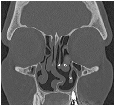 Figure 1. Coronal computed tomography demonstrates middle turbinate (a), secondary middle turbinate (b) and inferior turbinate (c). 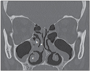 Figure 2. Sagittal computed tomography identifying right-sided middle turbinate (a), right-sided secondary middle turbinate (b) and right-sided inferior turbinate (c). 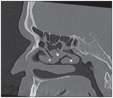 Figure 3. Coronal computed tomography demonstrates middle turbinate (a), secondary middle turbinate (b) and inferior turbinate (c). DISCUSSION The nasal turbinates are embryologically developed from lateral nasal wall excrescences which posteriorly form six crests, some of which fuse with each other, giving origin, among other structures, to the middle and superior turbinates. Because of the complexity in their development, innumerable anatomical variations may occur, middle turbinate pneumatization being the most frequent one. However, other rare variations may also be found, like in the case of secondary middle turbinate, a condition firstly reported by Khanobthamchai et al.(3), with a frequency of 1.5%, and that in other studies has ranged between 0.8% and 14.3%. Secondary middle turbinate is a bone structure covered by a tissue with soft-tissue density, originating from the lateral wall of the middle meatus, just below the basal lamella(4). Initially, it was described as a bilateral structure, but some authors have reported cases of unilateral involvement(5), leading to the questioning about the possibility of an underdiagnosed variation, probably confused with other conditions such as nasal polyps. Generally such variation is not related to osteomeatal obstruction and, therefore, it is not usually considered to be a predisponent factor of sinusitis. However, according to studies developed by El-Shazly et al.(6), complaints about frontal headache in association with sensation of nasal obstruction were frequently observed in 92.8% of their patients with such a variation. Also, the finding of other anatomical variations in association with secondary middle turbinate is frequent. According to a recent study(6), concomitant anomalies were found in 49.7 % of the evaluated study sample, and ethmoid bulla was found in 9% of such cases. Accessory turbinate is an anatomical variation from which secondary middle turbinate must be differentiated(6). Accessory turbinate is a medial and anterior extension from the uncinate process. For the differential diagnosis based on tomographic or even endoscopic images, it is important to observe the uncinate process(5), like in the present case where, in spite of the medial displacement of the uncinate process, making its visualization more difficult, it could be tomographically differentiated from the accessory turbinate. REFERENCES 1. Souza RP, Brito Júnior JP, Tornin OS, et al. Complexo nasossinusal: anatomia radiológica. Radiol Bras. 2006;39:367-72. 2. Eweiss A, Khatwa MMA, Zeitoun H. Trifurcate middle turbinate; an unusual anatomical variation. Rhinology. 2008;46:246-8. 3. Khanobthamchai K, Shankar L, Hawke M, et al. The secondary middle turbinate. J Otolaryngol. 1991;20:412-3. 4. Aouad RK, Strong EB. Secondary middle turbinate. Otolaryngol Head Neck Surg. 2010;142:140-1. 5. Nebil ARK, Aktas D, Yilmaz T, et al. Unilateral secondary middle turbinate with sinusitis. KBB-Forum. 2009;8:44-5. 6. El-Shazly AE, Poirrier AL, Cabay J, et al. Anatomical variations of the lateral nasal wall: the secondary and accessory middle turbinates. Clin Anat. 2012;25:340-6. 1. MDs, Trainees in Radiology and Imaging Diagnosis, Hospital Mater Dei, Belo Horizonte, MG, Brazil. 2. MDs, Radiologists, Service of Radiology, Hospital Mater Dei, Belo Horizonte, MG, Brazil. 3. MD, Radiologist, Chief of Service of Radiology, Hospital Mater Dei, Belo Horizonte, MG, Brazil. Mailing Address: Dra. Bruna de Oliveira Melim Aburjeli Rua Aimorés, 2255/1102, Bairro Lourdes Belo Horizonte,MG, Brazil, 30140-072 E-mail: bruninha86@hotmail.com Received June 4, 2012. Accepted after revision July 25, 2012. * Study developed at Hospital Mater Del, Belo Horizonte, MG, Brazil. |
|
Av. Paulista, 37 - 7° andar - Conj. 71 - CEP 01311-902 - São Paulo - SP - Brazil - Phone: (11) 3372-4544 - Fax: (11) 3372-4554