Radiologia Brasileira - Publicação Científica Oficial do Colégio Brasileiro de Radiologia
AMB - Associação Médica Brasileira CNA - Comissão Nacional de Acreditação
 Vol. 43 nº 4 - July / Aug. of 2010
|
ORIGINAL ARTICLE
|
|
Radiation dose optimization in routine computed tomography: a study of feasibility in a University Hospital |
|
|
Autho(rs): Juciléia Dalmazo1; Jorge Elias Júnior2; Marco Aurélio Corte Brocchi3; Paulo Roberto Costa4; Paulo Mazzoncini de Azevedo-Marques5 |
|
|
Keywords: Computed tomography; Radiation dose reduction; ALARA; Signal-to-noise ratio; CTDI; Optimization. |
|
|
Abstract: INTRODUCTION
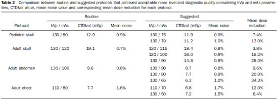
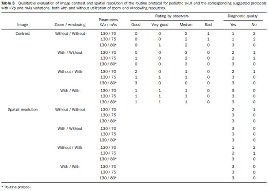
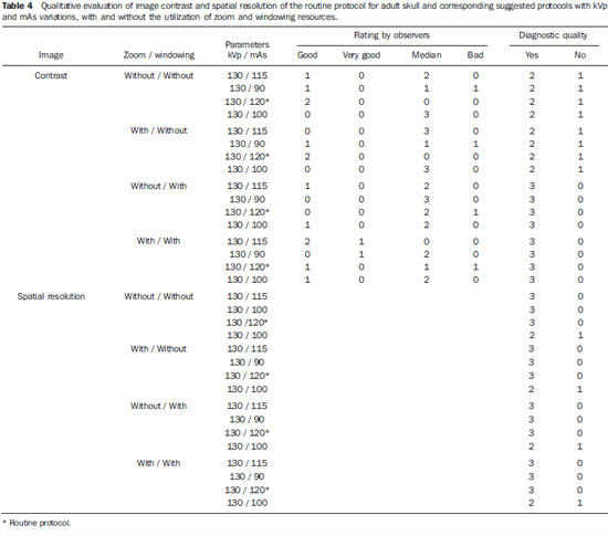
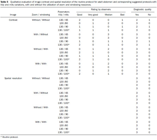
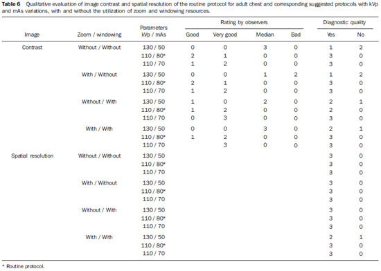
The annual number of computed tomography studies (CT) has constantly increased since its introduction in the clinical practice(1,2). Various factors contribute to this growth, including technological hardware improvements, leading to faster data acquisition with significant reduction in the images acquisition time, as well as the increase in the number of clinical indications for CT, associated to a greater availability of CT installed units and a relative tendency to costs reduction(3,4). The increasing utilization of diagnostic imaging methods employing ionizing radiation, particularly CT, is the main cause for the marked increase in the mean medical radiation dose per capita per year(2). Currently, the annual per capita radiation dose considered as secondary to medical care, particularly for diagnosis purposes, has overcome the dose received from environmental factors (food, radon gas and others)(1). As a result, there is an increasing concern of the medical community, equipment manufactures and even patients with the control of doses determined by the different diagnostic methods that rely on ionizing radiation(5,6). Besides occupational radiation protection, the clinical practice adopts the ALARA (As Low As Reasonably Achievable) principle as a guideline for the rational use of these imaging methods(3,4,7,8). Specifically considering the pediatric population, it is important to highlight that children present a considerably higher risk for development of radiation related neoplasia as compared with the adult population(1,9,10). Such higher risk is explained by the presence of a greater cell population undergoing division in the different organs and tissues in development, and also for the greater life expectancy both in absolute and relative terms. As an example, a oneyear-old child presents a 10 to 15 times higher risk than a 50-year-old adult for developing a malignant neoplasm, for the same radiation dose(1). For these reasons, there is an increasing concern with the radiation dose utilized in pediatric radiological studies, and particularly in the case of CT scans. Several published studies report strategies and actions to reduce radiation dose(3,5,9,11-13). There are several strategies in development or already in use in more modern equipment, such as, for example, tube current modulation according to the variation in the slice thickness for the evaluated anatomic region(6,14). However, in Brazil there are many relatively old CT systems in use with no software or parameter manipulation capabilities, which ultimately influence the dose delivered at each exam. Therefore, the present study is aimed to evaluate the feasibility of an optimization strategy to reduce radiation dose in single-slice helical CT protocols in a University Hospital. MATERIALS AND METHODS Strategy description Initially, data on the absorbed radiation dose in routine protocols for both adult and pediatric skull, chest and abdomen CT studies were collected. With such data, an evaluation of the impact of variations in the voltage parameters (kVp) and current vs. time (mAs) was undertaken considering the radiation dose and image quality, the latter studied by the noise measurement and subjective analysis of images obtained with specific phantoms. Equipment The tests were performed with a Somatom Emotion single-slice helical computed tomography unit (Siemens Medical Solutions; Erlangen, Germany). For the radiation dose measurements, cylindrical 15cm long polymethylmetacrylate phantoms were utilized, one with 32 cm in diameter, representing the torso, and another with 16 cm in diameter, representing the head (Figure 1), according to the American Association of Physicists in Medicine (AAPM) specifications. Such phantoms presented absorption and scattering characteristics that are similar to those of the anatomic structures of the skull and of the torso with holes in the center and at predetermined locations, at 1cm from the periphery at the 12, 3, 6 and 9 o'clock positions, allowing the insertion of ionization chambers. 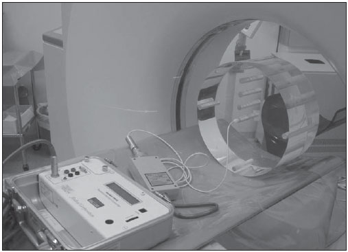 Figure 1. Electrometer, ionization chamber and body phantom exposed to the tomographic beam for CTDI calculation. The radiation dose measurements were obtained by means of a pencil type ionization chamber model 10X5-3CT (Radcal Corporation; Monrovia, CA, USA), coupled with a Radcal electrometer model 9015. Figure 1 shows the assembly for absorbed dose measurement for the standard body phantom. The images for the quality study with the proposed parameter variations were obtained by utilizing the specific AAPM standard phantom, model 76-410 which was freely borrowed by Instituto de Eletrotécnica e Energia da Universidade de São Paulo (IEE-USP)(Figure 2). 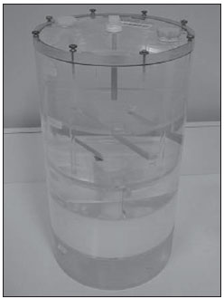 Figure 2. Quality phantom for analysis of noise, contrast and spatial resolution of the image. Studied variables The kVp and mAs values were duly noted for each protocol, as well as their suggested variations for protocols that were developed for radiation dose reduction. The initial evaluation of radiation dose in routine protocols as well as the CT equipment calibration were performed by means of the correlation between the CTDIvol obtained with the ionization chamber positioned within the phantoms, considering the acquisition of a section at the central position of the chamber, and the CTDIvol provided by the equipment. The CTDIvol is obtained from the ratio between the weighted computed tomography dose index (CTDIw)(15,16) and the pitch, which is defined as the distance travelled by the table and one rotation of the X-ray source, divided by the total collimated beam width. The CTDIvol is referenced on the equipment by the DICOM tag (0018,9345). Considering that the index of correlation between the values obtained in the measurements performed with the ionization chamber and those provided by the equipment was practically 100% (r=0.99; p < 0.0001), the equipment CTDIvol measurements were selected for results presentation of and discussion. Strategy for the proposal of changes in routine CT studies protocols Based on the initial standard protocols, the variations of the kVp and mAs parameters applied to the tube were proposed, with the remaining parameters (slice thickness, pitch, pixel size and total exposure) being kept constant for each protocol, as shown on Table 1 for the protocol of skull CT. The CTDIvol was measured again for each proposed parameter change (Table 1). 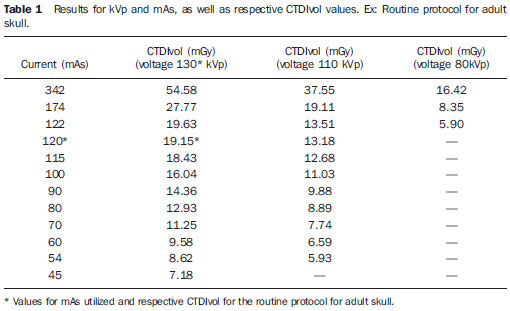 Evaluation of image quality The images quality was quantitatively evaluated by the measurement of quantic noise, and qualitatively by the subjective evaluation of images obtained in the AAPM standard phantom, independently and blindly performed by three radiologists with more than 10 years of experience. The quantic noise is the result of the variation in the number of X-ray photons absorbed by the detector in a determined time interval and, considering the geometric characteristics of the image (pixel size, matrix size, slice thickness) as fixed, presents an inversely proportional relation with the dose received by the patient. The methodology adopted for the evaluation of images quality was based on the Agência Nacional de Vigilância Sanitária (ANVISA - Brazilian agency of sanitary vigilance) guidelines "Medical radiodiagnosis: safety and equipment performance" that establishes the practical aspects for the standards established by Ordinance 453 of the Brazilian Ministry of Health(17), considering the quantic noise as the standard deviation of the values for the gray scale for a central square 5 × 5 mm region on a uniform image (Figure 3) divided by the nominal pixel value of that region. Although the ANVISA protocol indicates the need to evaluate the noise at five different points, as it refers to medical equipment quality control procedures, in the present study the option was made to simplify such procedure, by performing the measurement in the central region of the phantom for quality evaluation so as to observe the noise variation considering a situation of greater attenuation and beam hardening. The noise on the digital image was evaluated by means of the public domain ImageJ software(18), considering as acceptable those values < 1%(9). 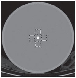 Figure 3. CT section of phantom for quality study, with four selected central areas selected in the ImageJ software for image noise calculation. The qualitative analysis was performed for the tested protocols that presented the lowest dose rates and with noise levels within the established limit. The evaluation comprised spatial resolution and high resolution contrast tests performed by the radiologists utilizing the ImageJ software without and with the use of windowing and zoom resources. The visualized objects indicated by the radiologists on the images were compared with the test objects map included in the phantom (Figure 4). 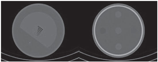 Figure 4. CT sections of phantom for quality study and analysis of image spatial resolution and contrast. The images were evaluated separately and in duets by means of a questionnaire with nine questions for each set of images, aimed at the study of images contrast and resolution threshold, considering even what set of objects were visible for the diagnosis of an image. The observer classified whether the image had diagnostic quality or not, considering the visibility of existing objects. Additionally, a classification of the image quality was requested, considering the values 1(very good), 2(good), 3(median), 4(bad) and 5(very bad). RESULTS The CTDIvol and mean noise values obtained for the routine protocols of pediatric skull, adult skull, abdomen and chest CT are presented on Table 2. Mean noise of 1.6% was observed for the chest protocol. By utilizing fixed 80, 100 and 130 kVp values, measurements of CTDIvol and mean noise were performed in 22 variations of the pediatric skull protocol (mAs ranging from 45 to 271), 26 variations of the adult skull protocol (mAs ranging from 45 to 342), 28 variations of the adult abdomen protocol (mAs ranging from 24 to 182) and 18 variations of the chest protocol (mAs ranging from 29 to 100). Based on these results, new protocols were suggested considering the combination of lowest CTDIvol obtained in association with the defined noise threshold, which were utilized for the evaluation of the quality of the image obtained in the standard AAPM phantom specific for quality evaluation. The data on the suggested protocols and respective CTDIvol and mean noise values, as well as the average dose reduction comparatively with the routine protocols, are presented on Table 2. As regards the qualitative evaluation of the suggested protocols, the specialists agreed that by using the zoom and windowing tools at the display monitor, the suggested protocols for pediatric skull, adult skull, adult abdomen and adult chest did not present any differences comparatively with the routine protocols in what concerns spatial resolution and contrast, as shown on Tables 3 to 6. However, it is important to note that in the case of adult skull, there was a trend towards improvement in the rating of the contrast quality of the images acquired with the suggested protocol, when the windowing and zoom tools were utilized. DISCUSSION The obtained results allowed the observation of an excellent correlation between the CTDIvol values obtained by measurements performed with the ionization chamber and those provided by the equipment, which makes it so much easier to know the actual radiation dose utilized in CT studies in general. Such high correlation, which was actually expected, confirms the appropriateness of the equipment calibration. The mean noise values for the routine protocols were within the established threshold of 1%, except for the chest protocol, in which the mean noise value was 1.6%. Such value is explained by the fact that the chest CT utilizes narrow collimation in order to obtain thin slices, thus determining a smaller quantity of photons incident on the detectors. For this reason, there was no proposal for dose reduction in the chest protocols, as the noise level was already above the established limit. By observing the quantitative and qualitative evaluation of the images, it becomes clear that all CT protocols could utilize reduced kVp and mAs parameters while maintaining diagnostic quality, and determining a lower radiation dose in each study, a fact that is in agreement with other studies in the literature(19-23), particularly with respect to mAs reduction. Such a reduction is particularly significant for CT studies in children, which has been a constant preoccupation over the last decade(10,13,24,25). With the methodology utilized, the authors observed a reduction in radiation dose between 3.8% and 34.3%. It is possible to achieve further radiation dose reduction with techniques that take anthropometric individual data into consideration, as reported by Kalra et al.(22), or by working with less conservative noise levels, for example in the order of 5%. It should be reminded that any level of radiation dose reduction must be pursued as determined by the ALARA principle(3,4,7,8). Also, it must be highlighted that in practically all subjective evaluations performed by the radiologists, better scores were observed with the utilization of the zoom and windowing resources. Such results corroborate findings reported in the literature discussing the relation between reduction of radiation dose in CT studies, image quality and reliability of the subjective evaluation(26). Considering that in digital imaging method the processes of images acquisition and display are separated, it is possible to independently optimize each process. Thus, within determined limits, it is possible to compensate for a loss in contrast due to a decrease in the signal/noise ratio by utilizing easily manipulated tools. The optimization of contrast resolution, with the consequential potential for dose reduction, is the main advantage of digital technology as far as the patients radiological protection is concerned. The main limitation of the present study lies on the fact that the variations of other CT parameters such as pitch, slice thickness and rotation time were not approached. It is a known fact that when the pitch is increased the patient is exposed to a higher radiation dose. However such limitation is an expression of reality in several Brazilian CT centers, where limited resources do not allow an appropriate technological update of relatively old apparatuses as compared with units equipped with new resources for dose efficiency management, calibration of pediatric images quality, measurements of pediatric doses and protocols specifically designed for pediatrics(12). CONCLUSION Based on the results of the present study, the optimization of CT radiation doses in a university hospital was feasible with the proposed methodology utilizing phantoms and a pencil-type ionization chamber, achieving a radiation dose reduction of up to 34.3% for selected study protocols. REFERENCES 1. Picano E. Sustainability of medical imaging. BMJ. 2004;328:578-80. 2. Huda W. What ER radiologists need to know about radiation risks. Emerg Radiol. 2009;16:335-41. 3. Semelka RC, Armao DM, Elias J Jr, et al. Imaging strategies to reduce the risk of radiation in CT studies, including selective substitution with MRI. J Magn Reson Imaging. 2007;25:900-9. 4. Frush DP, Donnelly LF, Rosen NS. Computed tomography and radiation risks: what pediatric health care providers should know. Pediatrics. 2003;112:951-7. 5. Picano E, Vano E, Semelka R, et al. The American College of Radiology white paper on radiation dose in medicine:deep impact on the practice of cardiovascular imaging. Cardiovasc Ultrasound. 2007;5:37. 6. Kalender WA, Buchenau S, Deak P, et al. Technical approaches to the optimisation of CT. Phys Med. 2008;24:71-9. 7. ICRP. 1990 Recommendations of the International Commission on Radiological Protection. ICRP Publication no. 60. Oxford, UK: Pergamon; 1991. 8. Kalra MK, Maher MM, Toth TL, et al. Strategies for CT radiation dose optimization. Radiology. 2004;230:619-28. 9. Verdun FR, Lepori D, Monnin P, et al. Management of patient dose and image noise in routine pediatric CT abdominal examinations. Eur Radiol. 2004;14:835-41. 10. Donnelly LF. Reducing radiation dose associated with pediatric CT by decreasing unnecessary examinations. AJR Am J Roentgenol. 2005;184:655-7. 11. Berdon WE, Slovis TL. Where we are since ALARA and the series of articles on CT dose in children and risk of long-term cancers: what has changed? Pediatr Radiol. 2002;32:699. 12. Morgan HT. Dose reduction for CT pediatric imaging. Pediatr Radiol. 2002;32:724-8; discussion 751-4. 13. Siegel MJ, Schmidt B, Bradley D, et al. Radiation dose and image quality in pediatric CT: effect of technical factors and phantom size and shape. Radiology. 2004;233:515-22. 14. McCollough CH, Bruesewitz MR, Kofler JM Jr. CT dose reduction and dose management tools: overview of available options. Radiographics. 2006;26:503-12. 15. Gerber TC, Kuzo RS, Morin RL. Techniques and parameters for estimating radiation exposure and dose in cardiac computed tomography. Int J Cardiovasc Imaging. 2005;21:165-76. 16. Pina DR, Duarte SB, Ghilardi Netto T, et al. Controle de qualidade e dosimetria em equipamentos de tomografia computadorizada. Radiol Bras. 2009;42:171-7. 17. Brasil. Ministério da Saúde. Secretaria de Vigilância Sanitária. Portaria nº 453, de 01 de junho de 1998. Brasília: Diário Oficial da União; 02/06/1998. 18. Abramoff MD, Magelhaes PJ, Ram SJ. Image processing with ImageJ. Biophotonics International. 2004;11:36-42. 19. Mayo JR, Hartman TE, Lee KS, et al. CT of the chest: minimal tube current required for good image quality with the least radiation dose. AJR Am J Roentgenol. 1995;164:603-7. 20. Heyer CM, Mohr PS, Lemburg SP, et al. Image quality and radiation exposure at pulmonary CT angiography with 100- or 120-kVp protocol: prospective randomized study. Radiology. 2007;245:577-83. 21. Cohnen M, Fischer H, Hamacher J, et al. CT of the head by use of reduced current and kilovoltage: relationship between image quality and dose reduction. AJNR Am J Neuroradiol. 2000;21:1654-60. 22. Kalra MK, Prasad S, Saini S, et al. Clinical comparison of standard-dose and 50% reduced-dose abdominal CT: effect on image quality. AJR Am J Roentgenol. 2002;179:1101-6. 23. Jung KJ, Lee KS, Kim SY, et al. Low-dose, volumetric helical CT: image quality, radiation dose, and usefulness for evaluation of bronchiectasis. Invest Radiol. 2000;35:557-63. 24. Brenner D, Elliston C, Hall E, et al. Estimated risks of radiation-induced fatal cancer from pediatric CT. AJR Am J Roentgenol. 2001;176:289-96. 25. Donnelly LF, Emery KH, Brody AS, et al. Minimizing radiation dose for pediatric body applications of single-detector helical CT: strategies at a large children's hospital. AJR Am J Roentgenol. 2001;176:303-6. 26. MacKenzie JD, Nazario-Larrieu J, Cai T, et al. Reduced-dose CT: effect on reader evaluation in detection of pulmonary embolism. AJR Am J Roentgenol. 2007;189:1371-9. 1. Physicist, Fellow PhD degree, Faculdade de Medicina de Ribeirão Preto da Universidade de São Paulo (FMRP-USP), Ribeirão Preto, SP, Brazil 2. MD, PhD, Professor and Coordinator at Centro de Ciências das Imagens e Física Médica (CCIFM) - Faculdade de Medicina de Ribeirão Preto da Universidade de São Paulo (FMRP-USP), Ribeirão Preto, SP, Brazil 3. Master, Physicist in Radiodiagnosis at Hospital das Clínicas da Faculdade de Medicina de Ribeirão Preto da Universidade de São Paulo (HCFMRP-USP), Ribeirão Preto, SP, Brazil 4. PhD, Professor at Department of Nuclear Physics, Instituto de Física da Universidade de São Paulo (IFUSP), São Paulo, SP, Brazil 5. PhD, Associate Professor at Centro de Ciências das Imagens e Física Médica (CCIFM), Department of Medical Practice, Faculdade de Medicina de Ribeirão Preto da Universidade de São Paulo (FMRP-USP), Ribeirão Preto, SP, Brazil Study developed at Centro de Ciências das Imagens e Física Médica (CCIFM) - Hospital das Clínicas da Faculdade de Medicina de Ribeirão Preto da Universidade de São Paulo (HCFMRP-USP), Ribeirão Preto, SP, Brazil Mailing address: Dr. Jorge Elias Júnior Centro de Ciências das Imagens e Física Médica, HCFMRP-USP Avenida Bandeirantes, 3900, Monte Alegre 14048-090. Ribeirão Preto, SP, Brazil Email: jejunior@fmrp.usp.br Received December 9, 2009 Accepted after revision June 1st, 2010 |
|
Av. Paulista, 37 - 7° andar - Conj. 71 - CEP 01311-902 - São Paulo - SP - Brazil - Phone: (11) 3372-4544 - Fax: (11) 3372-4554
