Radiologia Brasileira - Publicação Científica Oficial do Colégio Brasileiro de Radiologia
AMB - Associação Médica Brasileira CNA - Comissão Nacional de Acreditação
 Vol. 40 nº 6 - Nov. / Dec. of 2007
|
ICONOGRAPHIC ESSAY
|
|
Esophageal motility: an iconographic essay on dynamic esophageal scintigraphy |
|
|
Autho(rs): Maria Expósito Penas |
|
|
Keywords: Esophageal scintigraphy, Transit time, Motility, Achalasia, Parameters |
|
|
Abstract:
Maria Expósito Penas PhD, Associate Professor, Department of Radiology - Nuclear Medicine at Faculty of Medicine, Universidade Federal do Rio de Janeiro (UFRJ), Rio de Janeiro, RJ, Brazil
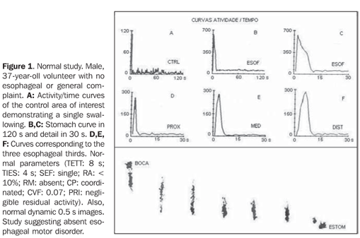
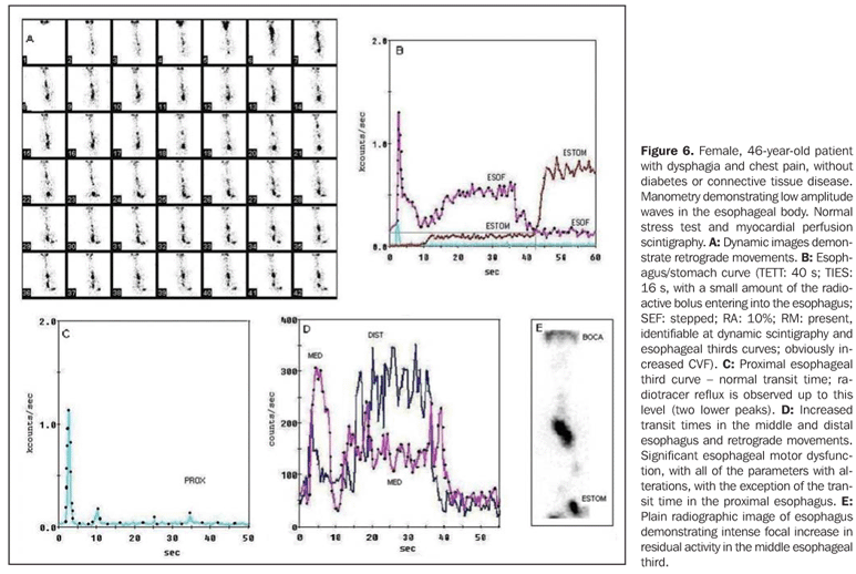
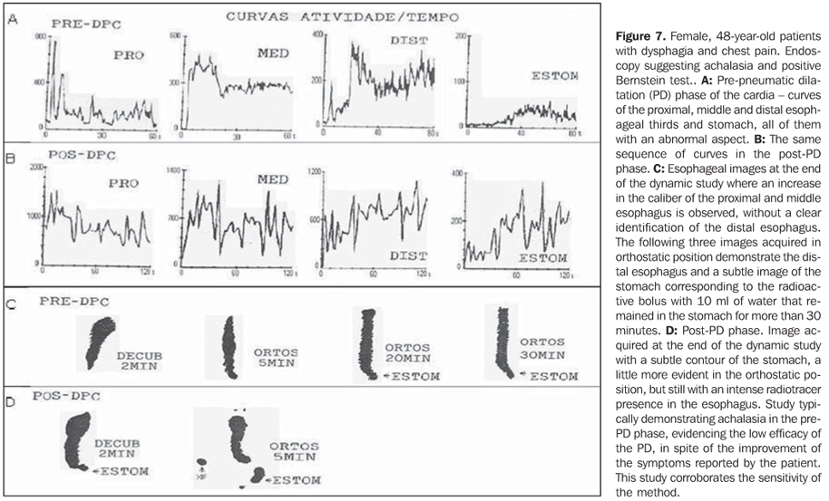
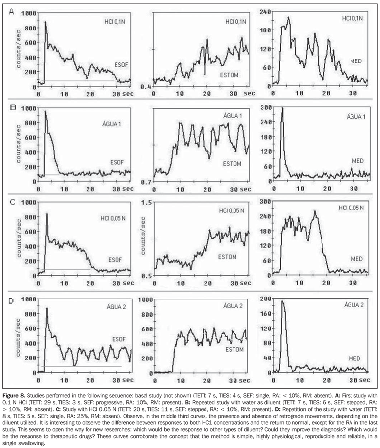
INTRODUCTION The present essay is aimed at showing an interesting selection of images and graphics demonstrating esophageal motility in a spectrum ranging from normal to the opposite extreme that is an advanced-stage achalasia. Besides studies utilizing room-temperature water for radiotracer dilution, further studies (Bernstein tests) were performed with volunteers (with free and informed consent) utilizing mild hydrochloric acid (at 0.1 N and 0.05 N concentration) to reproduce symptoms of gastroesophageal reflux. All of the studies were approached by a standard systematization including the following parameters (for normal values [n]): total esophageal transit time (TETT) (n: < 10.0 s), curve pattern (CP) (n: coordinated), residual activity (RA) (n: < 10%), time for initial entry into stomach (TIES) (n: < 6.0 s), stomach entry form (SEF) (n: abrupt), retrograde movements (RM) (n: absent), curve variation factor (CVF) (n: < 0.1), transit time in the proximal, middle and distal esophageal thirds (PTT, MTT, DTT) (respectively n: < 3.0 s, < 6.0 s and < 10.0 s) and a plain radiographic image of the esophagus (PRI) at the end of the dynamic scintigraphy (n: absent or mild residue)(1-3).
REFERENCES 1. Russell COH, Hill LD, Holmes ER III, Hull DA, Gannon R, Pope CE II. Radionuclide transit: a sensitive screening test for esophageal dysfunction. Gastroenterology 1981;80:887–892. [ ] 2. Blackwell JN, Hannan WJ, Adam RD, Heading RC. Radionuclide transit studies in the detection of oesophageal dysmotility. Gut 1983;24:421–426. [ ] 3. Penas ME, Orlando MMC, Koch HA. Dynamic esophageal scintigraphy parameters to analyze in single liquid bolus swallow. Alasbimn J 2006; 8(33). [ ]
Received March 8, 2007. Accepted after revision May 8, 2007.
* Study developed in the Department of Radiology - Nuclear Medicine at Faculty of Medicine, Universidade Federal do Rio de Janeiro (UFRJ), Rio de Janeiro, RJ, Brazil.
|
|

Av. Paulista, 37 - 7° andar - Conj. 71 - CEP 01311-902 - São Paulo - SP - Brazil - Phone: (11) 3372-4544 - Fax: (11) 3372-4554