Radiologia Brasileira - Publicação Científica Oficial do Colégio Brasileiro de Radiologia
AMB - Associação Médica Brasileira CNA - Comissão Nacional de Acreditação
 Vol. 40 nº 3 - May / June of 2007
|
ORIGINAL ARTICLE
|
|
The correlation between DEXA and CDEXA methods in bone mineral absorptiometry |
|
|
Autho(rs): Cláudio Gilberto Defavori, Gabriel Adrian Sarriés |
|
|
Keywords: Dual energy x-ray absorptiometry, Dual energy radiographic absorptiometry, Computed x-ray densitometry, Correlation, Interchangeability |
|
|
Abstract:
IMD, Radiologist at CPA Radiologia, Piracicaba, SP, Brazil
INTRODUCTION Dual energy x-ray absorptiometry (DEXA) is a technique widely utilized for measuring mass and bone mineral density (BMD). Computed dual energy conventional x-ray absorptiometry (CDEXA) is different from DEXA in the way x-rays absorption data acquisition is performed, but the mathematical algorithms utilized for absorptiometry data processing are similar. The CDEXA system present some interesting features as follows: higher spatial resolution and smaller area for locating the equipment as compared with DEXA systems; native images, allowing a radiological analysis to offer reliable densitometric data; lower total investment allowing a more disseminated and comprehensive use in the population; estimated radiation doses of 1.3 mSv and 2.6 mSv, respectively, for hip and spine assessment, and a probable increase in the diagnostic specificity, considering the feasibility of an individualized radiological analysis. It is necessary to know if results obtained for subjects with the CDEXA system may be reliably interchanged with results for the same subjects with a DEXA device, as previously done for comparison between absorptiometry techniques(1,2). The present study was aimed at comparing lumbar spine and femoral neck BMD measurements with these devices in a populational sample, regardless race/ethnicity, gender, age, body weight, or clinical status, to investigate the performance relation between both devices in a full-operational condition.
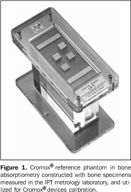
MATERIALS AND METHODS DEXA studies were performed with a Hologic® QDR 4500C apparatus, and CDEXA studies, with a Cromox® CXD 3.2.1 system, calibrated with a Cromox® reference phantom for bone absorptiometry. According to Blake et al.(3), the DEXA technology performs the scanning of the site in analysis, recording the x-ray beam power with dual kilovoltage components. This recording is performed by a solid-state sensor which detects the transmitted radiation and transfers the corresponding digital value into a computer, generating two image matrices corresponding to the two photon energies. The mathematical treatment of absorptiometry data is performed through the analysis of two transmission equations for each pixel of the double image matrix of the site of interest, one equation for each of the two x-ray beam kilovoltages. For each photon energy, an equation with exponential relation between attenuation and transmitted radiation energy is converted into a linear equation by application of a logarithmic function. Once the relation between the soft tissues attenuation coefficients for both energies is defined, and once the bone mineral component attenuation coefficients for both energies are known, the two resulting linear equations present only two unknown magnitudes corresponding to the densities per area of bone mineral component and soft tissues. The resolution of the linear algebric system defines the two densities per area on each pixel of the image matrix. The integration in area of density of the bone mineral component, followed by the division of the integrated value by the total area, results in the BMD arithmetic mean of the site evaluated. According to the manufacturer's information, the CDEXA technology also provides the two image matrices by exposure of the site analyzed by x-ray beams to two photon energies, through the operation of the bulb with two kilovoltages. The radiation beam energy is recorded on conventional radiographic films. Aiming at controlling variations both in the characteristics of the bulb emission and films chemical processing, calibration metal phantoms are submitted to radiological exposure simultaneously with the patient. The transference of absorptiometry data into the computer is made by means of radiographic films digitization by a scanner with solid-state sensors, which catch the luminosity transmitted and transfer the corresponding values into the computer, constructing two image matrices corresponding to the photon energies. The values of these image matrices are adjusted according to the phantoms response to radiation, so the absorptiometry data are corrected for a pattern of x-ray emission. The manufacturer informs that the mathematical treatment of the absorptiometry data is performed as previously described for the DEXA technique, until the BMD arithmetic mean for the site analyzed is obtained. According to Steel et al.(4), the effective radiation doses to the patient, in fan beam DEXA for studying hip and lumbar spine, are respectively 56 µSv and 59 µSv, and may reach 74,7 µSv, according to Maher(5). With the CDEXA technology, where the images acquisition is made on conventional radiographic films, the estimated effective radiation doses for the hip and lumbar spine assessment are respectively 1.3 mSv and 2.6 mSv. This estimation is made because the CDEXA technique requires two images from each site (generated in two kilovoltages), and the typical, individual effective dose for acquisition of lumbar spine images is 1.3 mSv, according to the U.S. Food and Drug Administration(6), resulting in an effective radiation dose of 2.6 mSv for lumbar spine evaluation. The dose required for generation of a hip image corresponds to 50% of the dose required for lumbar spine, so the total effective radiation dose for hip assessment is 1.3 mSv. The comparison between the absorptiometry techniques has taken into consideration the results of density/area in gram units of bone mineral component/cm² of the lumbar spine (L2 to L4 vertebrae) and right femoral neck. The anteroposterior projection was adopted for lumbar spine assessment; and for the proximal femur, the orthogonal projection (the usual positioning for bone densitometry) achieved by means of 30° internal rotation of the foot was adopted. According to the manufacturer's information, the Cromox® reference phantom for bone absorptiometry (Figure 1) includes four plates of oven-dried bovine bone specimens with regular thickness, whose mass and area were measured by the metrology laboratory of Instituto de Pesquisas Tecnológicas (IPT) in São Paulo, SP, Brazil. The specimens were kept in the oven for drying until their mass presented a variation of less than 1% along an eight-hour interval. The drying process was completed in 50 hours. The mass/area ratio provides a bone density in g/cm² for calibration of absorptiometry results.
Forty-three volunteers, with no distinction of race/ethnicity, gender, age, body weight or clinical status, who had accepted the terms of agreement to participate in the present study, were evaluated during the period between March/2003 and June/2003. The patients were included in the study regardless their clinical or physical status, since the study was aimed at comparing the performance of the two equipment units under adequate operational conditions for achievement of reliable results. For the right hip, the patients´ ages ranged between 48 and 84 years (mean and standard deviation = 63.0 ± 9.5), while for the lumbar spine, ages ranged between 48 and 79 years (mean and standard deviation = 60.4 ± 8.6). Forty-one pairs of results for right hip and 32 pairs for lumbar spine were obtained by the DEXA and CDEXA systems, however not all the patients contributed with results for both sites (hip and lumbar spine). The rejection criteria adopted for exclusion of pair of results of BMD measurements by both equipment units were: a) site thickness (abdominal or coxofemoral) > 24 cm; b) interference from soft tissues: excess of gas on the lumbar spine vertebrae or overlapping of abdominal fat and femoral neck; c) results clearly non-compatible with the radiological evaluation of the site, in any of the equipment units. Additionally, excess of gas on the Cromox® metal phantom, inherent to the CDEXA method, was considered as a rejection criterion. Once these criteria were applied, two pairs of results for hip affected by soft tissues interference (overlapping of abdominal fat and femoral neck), one pair of results clearly non-compatible with the radiological evaluation of the hip, and one pair of results of hip where the presence of excessive gas on the lumbar vertebrae was observed were excluded from the study, and the remaining 38 pairs of results for hip, and 32 pairs of results for lumbar spine were compared by regression analysis. However, some discrepancies in the positioning of the patients on both devices were observed (lack of strict observance of the 30° for internal rotation of the foot, as well as the lumbar spine alignment was not strictly reproduced). Despite the known influence of clothing elastic bands, feces and little abdominal gas on densitometry results, data from many patients have not been included since these items were not considered as rejection criteria. These occurrences may affect the correlation of results obtained by both equipment units. Montgomery's(7) regression and linear correlation techniques were utilized for statistical data analysis calculated by supplementary regression and correlation macros(8) with Microsoft Microsoft Excel and Statistic Analysis System (SAS) applications(9), and tested by Gauss and Markov mathematical models assumptions of independence, normality and null hypothesis rejection.
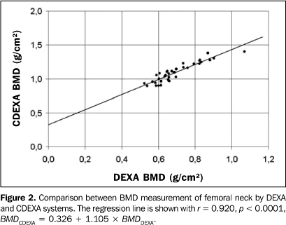
RESULTS The comparison between BMD measurements with both densitometry devices is shown on Figure 2 for the femoral neck, and Figure 3, for the lumbar spine.
For the femoral neck, the linear regression function for the group (n = 38, t-Student distribution) is:
where: BMDCDEXA and BMDDEXA are, respectively, the BMD values measured by CDEXA and DEXA; the correlation coefficient for femoral neck is r = 0.920, p < 0.0001. The data distribution and the regression line are shown on Figure 2. For the lumbar spine, the linear regression function for the group (n = 31, t-Student distribution) is:
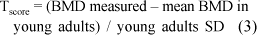
where the correlation coefficient is r = 0.923, p < 0.0001. The data distribution and the regression line are shown on Figure 3. In the clinical practice, the results expressed as Tscore are significant for evaluating bone densitometry studies, since the publication of the World Health Organization (WHO) Study Group report, according to Blake et al.(3). The Tscore adimensional parameter is defined by the equation: Tscore = (BMD measured – mean BMD in
where: young adults SD is the standard deviation for the young adults population. Even if there is a BMD correlation, Tscore measured in both systems are compatible only if the values for mean BMD in young adults and young adults SD are appropriate, these reference values for young adults for both systems being obtained from their respective results reports. The mean BMD calculation in young adults corresponds to the BMD/young adults percentage ratio in the site, while the young adults standard deviation corresponds to the BMD difference/mean BMD in young adults/Tscore ratio in the site. For each site, the value obtained for each of these parameters is the respective arithmetic mean among the whole group results. The arithmetic mean of the Tscore difference (DTscore arithmetic mean) is calculated for the group, the DTscore adimensional parameter being defined as:
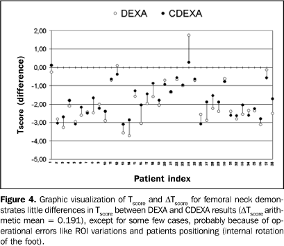
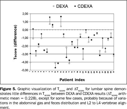
and calculated for each patient, resulting in a DTscore arithmetic mean = 0.019l for the femoral neck and DTscore arithmetic mean = 0.228 for the lumbar spine. The DTscore graphic representation is shown on Figure 4 for the femoral neck and Figure 5 for the lumbar spine, where the patients are indexed 1 to 38 (hip group), and 1 to 31 (lumbar spine group).
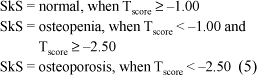
Aiming at investigating how the results provided by both devices can agree as regards the classification of the patient's skeletal status according to the WHO criteria, as normal, osteopenia and osteoporosis, as reported by Blake et al.(3), the results for each site were compared through the skeletal status parameter (SkS):
Agreement as regards skeletal status is defined as the same skeletal status classification for results obtained by both systems for a same patient, according to the mentioned mathematical criterion. Based on this definition, agreement as regards skeletal status was found in 76.3% of pairs of results for hips (n = 38), and in 77.4% for lumbar spine (n = 31). It is important to note that some pairs of BMD results present differences of decimals or centesimals of Tscore unit, but they are sufficient for determining different skeletal status. Another alternative method for evaluating agreement as regards skeletal status is considering the results of a same skeletal status obtained in both devices for a same patient and for at least one of the sites evaluated. In this case, agreement as regards skeletal status was found in 96.2% of pairs of results for hips and/or lumbar spine of the 26 patients submitted to measurements of both sites.
DISCUSSION BMD results were linearly correlated with r = 0.920 for femoral neck and r = 0.923 for lumbar spine, similarly to the correlation coefficient r ³ 0,95 reported by Faulkner et al.(10) in a comparison between DEXA Lunar and Hologic systems. Statistically, a correlation coefficient > 0.9 is very significant, expressing a small dispersion for results obtained in both devices for the same patients. Therefore, measurements performed in a device may be obtained in the other with small mean statistical differences. The correlation occurs at the same level both for hip and lumbar spine, since the correlation coefficients for both sites are very similar. This means that the significant correlation between the two methods is non-specific for the other site. The DTscore arithmetic means for hip and lumbar spine are very low, and not statistically significant, since these values correspond to a BMD variation lower than the least significant change (LSC) which is the lower BMD variation considered as statistically significant according to the International Society for Clinical Densitometry (ISCD). Using the algorithm offered by the ISCD(11), the LSC is determined for each device and for each site. For the CDEXA device, with 30 pairs of results, the LSCs were 0.088 g/cm² for hip, and 0.123 g/cm² for lumbar spine, resulting in DTscore LSC = 0.39 for hip, and DTscore LSC = 0.62 for lumbar spine., according to the equation:
According to the same algorithm, the LSC corresponds to 2.77 times the variance coefficient (VC) at 68% confidence level. for the DEXA system, VC = 1.8% is reported by Blake et al.(3), allowing the calculation of the LSC for each site, resulting in DTscore LSC = 0.45 for the hip, and DTscore LSC = 0.49 for the lumbar spine, by means of the equation (6). The DTscore arithmetic mean = 0.191 for the femoral neck and DTscore arithmetic mean = 0.228 for the lumbar spine are lower than DTscore LSC values for these sites in both systems, that is to say, they are sufficiently low to consider as adequate the interchangeability between Tscore results found through both systems. Figures 4 and 5 show constant, very little differences in Tscore, except for some few cases of patients with indices 14, 16, 24 and 27 for hip, and patients with indices 2,12,14, 20 and 27 for lumbar spine. For these cases, the not so little differences in Tscore probably occur as a result of operational errors such as variation in the definition of regions of interest (ROIs) and patient positioning (internal rotation of the foot, for the hip), variations in abdominal gas and feces distribution, and vertebrae L2 to L4 alignment (for the lumbar spine). In CDEXA devices, the inaccuracy of lumbar spine results because of the presence of abdominal gas and feces may be minimized if the images are rejected upon visual inspection of the radiographs, and the patient is submitted to a new radiographic exposure after appropriate intestinal preparation. The skeletal status parameter is introduced for the understanding of the interchangeability between the two methods in the clinical practice, since the first clinical interpretation of densitometry results is performed according to the WHO definitions. Considering the strictness of the WHO classification as regards status transition (normal/osteopenia, Tscore = –1.00; osteopenia/osteoporosis, Tscore = –2.50), an eventually non-elevated level of agreement is expected, even in comparisons between results from the same device, as a function of results deviation because of the device accuracy. With results classified according to the WHO categorization for each site, agreement as regards skeletal status does not occur for 24% of patients who had their hips evaluated, and for 23% of patients who had their lumbar spine evaluated. The agreement rate increases as the results are non-specifically classified for each site, i.e., considering as agreement as regards skeletal status the agreement in at least one of the two sites evaluated. Agreement as regards skeletal status is not found in only 4% of the patients evaluated by both systems when at least one of the two sites (hip or lumbar spine) is considered. For conversion of BMD values measured by CDEXA into DEXA scale, the appropriate equation obtained by linear regression for the femoral neck group is:
with p < 0.0001, while for the lumbar spine group the appropriate equation is:
with p < 0.0001. However, there is a necessity for additional studies involving a higher number of cases and with a more strict control over patients positioning and definition of ROI in the software for BMD measurement by both equipment units to evaluate the correlation between the devices when submitted to tests with a higher reproducibility of operational procedures. Acknowledgments The authors thank the manufacturers of the Cromox® CXD 3.2.1 system for providing technical information and license for utilization of the software during the study, allowing the processing of paired densitometric studies and those performed in the Hologic® QDR 4500C system owned by the author. The present study has been developed as a previous condition for the evaluation and acquisition of the Cromox® CXD 3.2.1 system by the author.
REFERENCES 1. Blake G, Rodin A, Fogelman I. A comparative study of dual photon absorptiometry and dual energy X-ray absorptiometry. J Bone Mineral Res 1989;4 Suppl. 1:S398. [ ] 2. Adachi JD, Webber CE. The interchangeability of radioisotope and X-ray based measurements of bone mineral density. Br J Radiol 1991;64:217–220. [ ] 3. Blake GM, Wahner HW, Fogelman I. The evaluation of osteoporosis: dual energy X-ray absorptiometry and ultrasound in clinical practice. 2nd ed. London, UK: Martin Dunitz, 1999. [ ] 4. Steel SA, Baker AJ, Saunderson JR. An assessment of the radiation dose to patients and staff from a Lunar Expert-XL fan beam densitometer. Physiol Meas 1998;19:17–26. [ ] 5. Maher KP. Department of Medical Radiations Science, RMIT University, Australia. [cited 2006 June 5]. Available in: http://homepage.mac.com/kieranmaher/digrad/DRPapers /DEXA–Dosimetry.html [ ] 6. U.S. Food and Drug Administration, Center for Devices and Radiological Health. Whole body scanning using computed tomography (CT). What are the radiation risks from CT? [cited 2006 June 5]. Availabble from: http://www.fda.gov/cdrh/ct/risks.html [ ] 7. Montgomery DC. Introduction to statistical quality control. New York: John Wiley & Sons, 1991. [ ] 8. McFredries P. Excel 5 super book. Rio de Janeiro: Berkeley Brasil Editora, 1994. [ ] 9. SAS Institute Inc. SAS/STAT guide for personal computers. 6th ed. Cary: SAS Institute, 1996. [ ] 10. Faulkner KG, Roberts LA, McClung MR. Discrepancies in normative data between Lunar and Hologic DXA systems. Osteoporos Int 1996;6: 432–436. [ ] 11. The International Society for Clinical Densitometry. Cálculo de precisão para densitometria óssea. Versão 2.1. [cited 2005 June 15]. Available from: http://www.iscd.org/visitors/xls/Precision CalcPortugueseRecommeded.xls [ ]
Received November 9, 2005.
* Study developed at CPA Radiologia, Piracicaba Unit, Piracicaba, SP, Brazil. |
|


Av. Paulista, 37 - 7° andar - Conj. 71 - CEP 01311-902 - São Paulo - SP - Brazil - Phone: (11) 3372-4544 - Fax: (11) 3372-4554