Radiologia Brasileira - Publicação Científica Oficial do Colégio Brasileiro de Radiologia
AMB - Associação Médica Brasileira CNA - Comissão Nacional de Acreditação
 Vol. 40 nº 3 - May / June of 2007
|
ORIGINAL ARTICLE
|
|
Magnetic resonance imaging for diagnosis of the chondral, meniscal and cruciate ligaments injuries of the knee |
|
|
Autho(rs): Francisco Consoli Karam, Jefferson Luís Braga da Silva, Marcos William Fridman, Armando Abreu, Rodrigo Di Mare Arbo, Marcelo Abreu, José Francisco Vieira, Luiz Antônio Simões Pires |
|
|
Keywords: Arthroscopy, Magnetic resonance imaging, Knee, Lesions, Sensitivity, Specificity |
|
|
Abstract:
IPhD of Medicine and Sciences of Health, Traumatologist and Orthopedist in the Group of Knee at Pontifícia Universidade Católica do Rio Grande do Sul (PUCRS), Porto Alegre, RS, Brazil
INTRODUCTION Magnetic resonance imaging (MRI) has been considered as the imaging method of choice for evaluating knee joints, almost completely replacing arthrography in the last decade(1). This is due the fact that this technique represents a both non-invasive and accurate alternative to arthroscopy in the diagnosis of internal knee derangements(2). Notwithstanding the controversies about MRI cost-benefit ratio(3), it is important to note that the technological development has brought higher precision to this method, allowing a reduction in the need for diagnostic arthroscopies. The present study is aimed at investigating if MRI of the knee following the global protocol (without specific sequences for the different parts of the knee joint) reproduces the results found in the literature(2,4–8), and defining for which intra-articular structures this imaging method could be useful as a diagnostic tool. For this purpose, we performed a transversal study with a prospective collection of data from 72 patients submitted to MRI in a service of imaging diagnosis. Arthroscopy performed by a single surgeon was utilized as a comparative standard method.
MATERIALS AND METHODS Transversal study with prospective data collection performed in the period between January/2003 and December/2005, evaluating 72 patients submitted to MRI of the knee, whose findings were compared with those from subsequent arthroscopy of the knee considered as the standard method. The sample included 37 MRI of the right knee and 35 of the left knee of 65 men and seven women in the age range between 19 and 67 years (mean age, 35.4 years). Inclusion criteria were the following: patients of both sexes; age ³ 18 years (skeletal maturity); patients' complain of symptoms in the knee joint and previous history compatible with intra-articular disease; the patient has been examined by the main author of the present study who, after a standard physical examination of the knee and specific tests for lesions of intra-articular structures, has indicated arthroscopy and requested MRI as a complement for the clinical diagnosis; the patient has undergone arthroscopy in the period between one and 90 days after MRI, and has not experienced any episode of trauma in this period. Patients whose lesions could not be classified because of dubious description or interpretation of MRI reports have not been included in the present study. A GE Signa, 1.5 tesla MRI equipment was utilized in the present study. The standard protocol for investigation of the knee utilized in all of the patients included the following steps: positioning of the patient in dorsal decubitus with slight external rotation of the knee (about 5º); utilization of specific surface coil; 4 mm slice thickness at 0.4 mm intervals; small field of view (FOV) for resolution maximization (14 to 16 cm, depending on the size of the patient), 256 × 192 or 256 × 256 matrices; planes and sequences: sagittal T1, sagittal PD-T2, coronal PD-T2, axial gradient-echo, axial T2, and sagittal PD with thin sections to evaluate the anterior cruciate ligament. All the studies were blindly evaluated by a single radiologist who has collaborated in the present study. All of the arthroscopic evaluations were performed in a surgical center by the same orthopedist — main author of the present study with a 15-year experience in this type of procedure —, several of them also with the assistance of a colleague of Hospital São Lucas and collaborator in the present study with a 25-year experience in arthroscopy of the knee. Arthroscopies were performed through the classic antero-lateral and antero-medial parapatellar portals. After insertion of the arthroscope through the lateral parapatellar portal, a routine inspection was performed in the whole joint for evaluation of medial and lateral compartments (condyles, tibial plateaus and menisci), intercondyle (cruciate ligaments), and finally femoropatellar joint (patellar and synovial cartilages). Investigation through medial and lateral suprapatellar portals was performed as necessary. Results from this initial analysis where compared with the MRI findings described by the radiologist and a new inspection was made to search for any lesion detected by the MRI which may have been missed in the first inspection. Finally, lesions were surgically corrected as necessary. Videoarthroscopy was not performed in patients who had not an indication for surgical treatment (videosurgery), and, because of ethical reasons, this procedure could not be blindly performed, considering that the availability of a previous MRI obliges the orthopedist to confirm its findings. Lesions in the following sites were analyzed: both menisci (medial and lateral), cruciate ligaments (anterior and posterior), and five different cartilaginous — medial and lateral condyles, medial and lateral plateaus, and patella. Meniscal lesions were classified according to changes in Tyrrel et al.(9) and Stoller et al.(10) classifications as follows: grade 0 (absence of lesions, or grade 1 and 2 lesions of Stoller et al.), grade 1 (lesion in one side or grade 3 lesion of Stoller et al.) and grade 2 (lesion in both sides of acute and complex lesions). Ligamentous lesions were classified according Heron & Calvert(4) into grade 0 (absence of lesion or minimal extension) grade 1 (partial lesion), and grade 2 (complete lesion). Finally, chondral lesions also were classified according changes in Outerbridge(11) and Tyrrel et al.(9) classifications, as follows: grade 0 (absence of lesion), grade 1 (superficial lesion or grade 1 Outerbridge lesion), grade 2 (superficial or deep, partial lesion not reaching the subchondral bone, or Outerbridge's grades 2 and 3), and grade 3 (complete lesion up to the chondral bone or Outerbridge's grade 4). The present study was designed in compliance with the guidelines and rules on research in humans (Resolution 196/1996 of National Council of Health), and was approved by the Committee on Ethics in Research of Hospital São Lucas of Pontifícia Universidade Católica do Rio Grande do Sul, where arthroscopies were performed and the present study was developed. All the patients submitted to arthroscopy were given information and guidance on the procedure, its risks and benefits, and agreed to be submitted to the surgical treatment, besides agreeing that their data was utilized in the present study. Microsoft Excel 2002 was utilized in the statistical analysis for data tabulation and charts, and SPSS 11.5 version for the global data analysis. Interobserver agreement as regards lesions classification, sensitivity, specificity, positive and negative likelihood values were analyzed. The confidence interval was 95%.
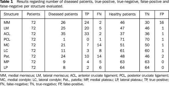
RESULTS Results regarding the number of diseased and healthy patients, true-positive and true-negative, and false-positive and false-negative diagnoses are shown in Table 1.
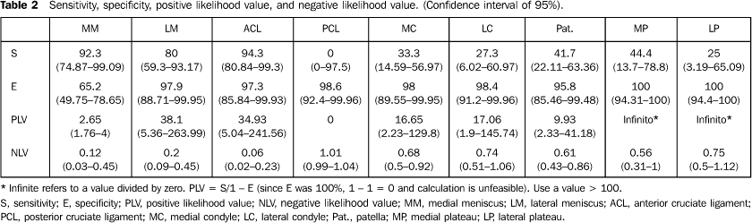
The kappa coefficient of agreement between MRI and arthroscopy as regards lesion classification was very good for lesions in the anterior cruciate ligament (0.84), good for lateral menisci (0.75), reasonable for medial menisci (0.50), and low for chondral lesions (< 0.5). MRI demonstrated high sensitivity for identifying anterior cruciate ligament tears (94%) and medial meniscal tears (92%), good sensitivity for lateral meniscal lesions (80%), and low sensitivity for all chondral lesions (< 50%) (Table 2). The positive likelihood value was near of above 10 for all the chondral lesions, anterior cruciate ligament lesions, and lateral meniscal lesions, and 2.65 for medial meniscal lesions (Table 2).
DISCUSSION As in the greatest majority of similar studies, arthroscopy was utilized as comparative standard method to evaluate the accuracy of MRI in the detection of intra-articular lesions of the knee(1,4,9,12,13). In the present transversal study with prospective data collection, a same team evaluated MRIs of 72 patients aiming at allowing the elaboration of a prior protocol including the methodology for analysis and classification of lesions. In the Brazilian literature, four studies are found with the same objective(8,14–16), only the last of them presenting an appropriate methodology, although not utilizing a classification for lesions or a prospective data collection. Similarly to many studies in the literature, the MRI system utilized was a 1.5 tesla equipment(4,17–19), the images acquisition protocol was the same for all the patients, and no special section of sequence for any particular anatomical structure of the knee could be added. The kappa agreement coefficient as regards the classification of the lesions was very good for the anterior cruciate ligament, good for the lateral meniscus, reasonable for the medial meniscus, and low for chondral lesions. It is important to note that this coefficient of agreement is very rigorous when compared with the number of correct diagnoses by the method over the total number of patients. These results would be better if we had quantified the error in percentage (weighted kappa coefficient), likewise Potter et al.(19) who, studying sequences specific for the joint cartilage, have defined four levels of discordance in classification of lesions, corresponding to 75% accuracy (1 level), 50% (2 levels), 25% (3 levels), and zero (complete discordance). Jee et al.(20) have already highlighted the relevance of classifying meniscal lesions and evaluating, by means of MRI, the level of stabilization or possibility of suture of these lesions, with consequent aid in the surgical planning and guidance for the patient on his postoperative course of treatment and recovery. The rationale is the same for chondral lesions, since the previous evaluation allows de surgeon to choose the most appropriate method of treatment, debridement or mosaicoplasty, for example. It is predictable that the future will require from MRI this level of performance. In the present study, rates of sensitivity, specificity and likelihood values (Table 2) were similar to those reported in the literature. Specificity was high (> 90%) for lateral meniscal lesions, medial and lateral condylar lesions, lesions in the posterior and anterior cruciate ligaments and in the medial and lateral tibial plateaus. For patellar lesions, specificity was very good, and merely good for medial meniscal lesions. The positive likelihood value was high for lesions in condyles, tibial plateaus, anterior cruciate ligaments and lateral meniscus, strongly indicating the presence of lesion, particularly when clinical studies point to the same direction. In the presence of a previous clinical suspicion, positive likelihood values > 10 strongly indicate the presence of disease(21). A merely reasonable specificity for medial meniscal lesions is related to the fact that some peripheral lesions may have healed in the period between the MRI and arthroscopy which ranged between one and 90 days, or due to the impossibility of visualizing peripheral lesions in the posterior horn of the medial meniscus, because of the difficult access in the arthroscopy, as suggested by Mackenzie et al.(22), with a resulting increase in the number of false-positive results. The negative likelihood value was low, especially for lesions in the medial meniscus and anterior cruciate ligament. In this case, de absence of lesion on the MRI study is a strong indicator that it really is not present, especially if the pretest probability is low. Because of the low prevalence of posterior cruciate ligament injury, i.e., only one patient affected among the 72 patients of the sample, an appropriate analysis of this structure was unfeasible. The lateral meniscus presented a good sensitivity and a negative likelihood value of 0.34, possibly contraindicating arthroscopy in the presence of poor clinical suspicion associated with a negative MRI. On the other hand, for chondral lesions, low sensitivity and negative likelihood value > 0.5 indicate that the absence of MRI findings does not exclude the presence of a lesion, added of the low efficacy of the physical examination for the diagnosis of these lesions. Spiers et al.(2) have already demonstrated the MRI low sensitivity and high specificity for chondral lesions, while contrast-enhanced MRI as well as arthrotomography seem to present higher accuracy, according to the literature(17). The utilization of specific protocols for visualization of cartilaginous tissues seems to considerably improve the MRI accuracy in the evaluation of these lesions. Potter et al.(19) have demonstrated that special MRI images for cartilages may result in considerable improvement in the accuracy of the method, since they have found a sensitivity of 87%, specificity of 94%, and accuracy of 92% in their study. In the present study, the patellar cartilage was the region where MRI presented the best performance, with reasonable sensitivity and a very good specificity, leading us to consider that, with the utilization of a special coil and a specific protocol, even without the use of contrast, MRI will significantly improve the diagnosis of patellar syndromes. Finally, it is important to note that mistakes may occur in the interpretation of diagnostic images, and so does it with MRI(23). This should be kept in mind, since MRI should not supersede the anamnesis and the physical examination, but should complement them in the search an accurate diagnosis. A diagnosis of intra-articular knee injury must be based on the aggregate of findings from anamnesis, physical examination (with specific tests for each structure), and analysis of radiological studies (plain radiographs, stress radiographs, arthrography, tomography and MRI).
CONCLUSIONS Magnetic resonance imaging is a useful tool in the clinical diagnosis of intra-articular knee injuries, as already demonstrated by similar results reported both in the Brazilian and international literature. MRI specificity, sensitivity, positive and negative likelihood values found for anterior cruciate ligament and meniscal injuries demonstrate the significant contribution of this method for the clinical diagnosis of such lesions. Considering the high positive likelihood value of the method, the visualization of a chondral lesion in a MRI study is a strong indicator of the presence of the disease. Since the negative likelihood value is not sufficiently low, the absence of an image of the lesion does not mean that such a lesion may be present.
REFERENCES 1. Oei EH, Nikken JJ, Verstijnen AC, Ginai AZ, Myriam Hunink MG. MR imaging of the menisci and cruciate ligaments: a systematic review. Radiology 2003;226:837–848. [ ] 2. Spiers AS, Meagher T, Ostlere SJ, Wilson DJ, Dodd CA. Can MRI of the knee affect arthroscopic practice? A prospective study of 58 patients. J Bone Joint Surg Br 1993;75:49–52. [ ] 3. Rangger C, Klestil T, Kathrein A, Inderster A, Hamid L. Influence of magnetic resonance imaging on indications for arthroscopy of the knee. Clin Orthop Relat Res 1996;330:133–142. [ ] 4. Heron CW, Calvert PT. Three-dimensional gradient-echo MR imaging of the knee: comparison with arthroscopy in 100 patients. Radiology 1992; 183:839–844. [ ] 5. Bui-Mansfield LT, Youngberg RA, Warme W, Pitcher JD, Nguyen PL. Potential cost savings of MR imaging obtained before arthroscopy of the knee: evaluation of 50 consecutive patients. AJR Am J Roentgenol 1997;168:913–918. [ ] 6. Jackson DW, Jennings LD, Maywood RM, Berger PE. Magnetic resonance imaging of the knee. Am J Sports Med 1988;16:29–38. [ ] 7. Brooks S, Morgan M. Accuracy of clinical diagnosis in knee arthroscopy. Ann R Coll Surg Engl 2002;84:265–268. [ ] 8. Vaz CE, Camargo OP, de Santana PJ, Valezi AC. Accuracy of magnetic resonance in identifying traumatic intraarticular knee lesions. Clinics 2005;60:445–450. [ ] 9. Tyrrell RL, Gluckert K, Pathria M, Modic MT. Fast three-dimensional MR imaging of the knee: comparison with arthroscopy. Radiology 1988; 166:865–872. [ ] 10. Stoller DW, Martin C, Crues JV, Kaplan L, Mink JH. Meniscal tears: pathologic correlation with MR imaging. Radiology 1987;163:731–735. [ ] 11. Outerbridge RE. The etiology of chondromalacia patellae. J Bone Joint Surg Br 1961;43:752–757. [ ] 12. Polly DW, Callaghan JJ, Sikes RA, McCabe JM, McMahon K, Savory CG. The accuracy of selective magnetic resonance imaging compared with the findings of arthroscopy of the knee. J Bone Joint Surg Am 1988;70:192–198. [ ] 13. Vande Berg BC, Malghem J, Poilvache P, Maldague B, Lecouvet FE. Meniscal tears with fragments displaced in notch and recesses of knee: MR imaging with arthroscopic comparison. Radiology 2005;234:842–850. [ ] 14. Schneider I, Schueda MA, Demore AB. Análise comparativa da ressonância magnética com a artroscopia no diagnóstico das lesões intra-articulares do joelho. Rev Bras Ortop 1996;31:373–376. [ ] 15. Severino NR, Camargo OP, Aihara T, et al. Comparação entre a ressonância magnética e a artroscopia no diagnóstico das lesões do joelho. Rev Bras Ortop 1997;32:275–278. [ ] 16. Yousef WJ, Thiele ES, Scuisato DL. Correlação diagnóstica da ressonância magnética com artroscopia nas lesões intra-articulares do joelho. Rev Bras Ortop 1999;34:375–380. [ ] 17. Gagliardi JA, Chung EM, Chandnani VP, et al. Detection and staging of chondromalacia patellae: relative efficacies of conventional MR imaging, MR arthrography, and CT arthrography. AJR Am J Roentgenol 1994;163:629–636. [ ] 18. Lundberg M, Odensten M, Thuomas KA, Messner K. The diagnostic validity of magnetic resonance imaging in acute knee injuries with hemarthrosis: a single-blinded evaluation in 69 patients using high-field MRI before arthroscopy. Int J Sports Med 1996;17:218–222. [ ] 19. Potter HG, Linklater JM, Allen AA, Hannafin JA, Haas SB. Magnetic resonance imaging of articular cartilage in the knee. An evaluation with use of fast-spin-echo imaging. J Bone Joint Surg Am 1998;80:1276–1284. [ ] 20. Jee WH, McCauley TR, Kim JM, et al. Meniscal tear configurations: categorization with MR imaging. AJR Am J Roentgenol 2003;180:93–97. [ ] 21. Deeks JJ, Altman DG. Diagnostic tests 4: likelihood ratios. BMJ 2004;329:168–169. [ ] 22. Mackenzie R, Keene GS, Lomas DJ, Dixon AK. Errors at knee magnetic resonance imaging: true or false? Br J Radiol 1995;68:1045–1051. [ ] 23. Muglia VF, Simão MN, Elias Jr J, Trad CS. Erros comuns de interpretação da ressonância magnética de joelho: como reconhecê-los e evitá-los. Radiol Bras 2001;34:161–166. [ ]
Received September 30, 2006.
* Study developed at Hospital São Lucas da Pontifícia Universidade Católica do Rio Grande do Sul (PUCRS), Porto Alegre, RS, Brazil. |
|

Av. Paulista, 37 - 7° andar - Conj. 71 - CEP 01311-902 - São Paulo - SP - Brazil - Phone: (11) 3372-4544 - Fax: (11) 3372-4554