Radiologia Brasileira - Publicação Científica Oficial do Colégio Brasileiro de Radiologia
AMB - Associação Médica Brasileira CNA - Comissão Nacional de Acreditação
 Vol. 41 nº 6 - Nov. / Dec. of 2008
|
ORIGINAL ARTICLE
|
|
Nomogram of fetal middle cerebral artery peak systolic velocity in a Brazilian population |
|
|
Autho(rs): Luciano Marcondes Machado Nardozza, Edward Araujo Junior, Christiane Simioni, Luiz Camano, Antonio Fernandes Moron |
|
|
Keywords: Middle cerebral artery, Doppler, Ultrasonography, Nomogram, Fetus |
|
|
Abstract:
IPhD, Associate Professor at Universidade Federal de São Paulo/Escola Paulista de Medicina (Unifesp/EPM), São Paulo, SP, Brazil
INTRODUCTION Red blood cells alloimmunization is characterized for being an immunological disorder resulting from the mother/fetus blood incompatibility, which determines a hemolytic disorder in the fetus and in the newborn. There is an excessively rapid red blood cells destruction, with consequential anemia, hyperbilirubinemia and severe, generalized edema. It is caused by specific antibodies produced by the mother and transplacentally transferred to the fetal bloodstream during pregnancy. After the discovery of the physiopathology, research into diagnosis techniques capable of measuring the anemia degree was started, in order to reduce perinatal morbidity and mortality rates. In 1961 Liley(1,) described the method of spectrophotometric amniotic fluid analysis in order to indirectly determine bilirubin concentration in the amniotic fluid and assess the hemolytic status, with the fluid being obtained by amniocentesis. However, amniocentesis is associated with complications such as early membrane rupture, preterm delivery, vaginal hemorrhage, infection and gestational loss in 0.5%(2). In the early eighties, with the discovery of high-resolution ultrasound-guided cordocentesis, this became the golden-standard method to assess the anemia degree in cases of alloimmunization during pregnancy(3). However, cordocentesis is associated with the worsening of maternal alloimmunization and fetal loss(4). In the late eighties, studies were undertaken using Doppler coupled with ultrasonography in an attempt to assess fetal hemodynamic parameters that could demonstrate the degree of fetal anemia(5,6). The main objective of these studies was determining an accurate method to avoid the risks of invasive procedures. In the mid-nineties, Mari et al.(7) demonstrated that the peak systolic velocity of the middle cerebral artery (MCA-PSV) was increased. Furthermore, they demonstrated that such vessel is easily visualized with an angle different from 0 degree, and that the measurement of this vessel presents low intra and interobserver variability(6). The same authors determined the first reference curve for MCA-PSV using multiples of the median(8). The present study was aimed at determining a reference curve for fetal MCA-PSV using multiples of the median, and also determining percentiles for this parameter in healthy pregnant women of the Brazilian population.
MATERIALS AND METHODS A cross sectional study was developed between August 2006 and August 2007, with 153 pregnant women assisted at the Unit of Fetal Medicine, Department of Obstetrics of Universidade Federal de São Paulo/Escola Paulista de Medicina (Unifesp/EPM), São Paulo, SP, Brazil. Ten of these pregnant women were excluded for the following reasons: five presented positive indirect Coombs test results, three did not present good technical conditions for the examination and two others presented chronic diseases (one with cardiopathy, the other with systemic lupus erythematosus). The present study was approved by the Committee for Ethics in Research of Unifesp/EPM, and the patients who agreed to voluntarily participate signed a term of free and informed consent. The present study evaluated 143 healthy pregnant women with single fetuses between 24 and 34 complete gestational weeks, with no malformation diagnosed in the prenatal period or confirmed in the postnatal period. The parameter utilized for determining the gestational age was the date of the last menstrual period (LMP) in the case of patients with normal periods. In the case of uncertainty about the LMP, the determination of gestational age was based on ultrasonography performed up to the 20th week. Gestational age limits were based on the criteria of fetal viability, this period being critical for the birth of premature fetuses which could potentially benefit from an appropriate evaluation of the vascular flow. All the studies were performed by a single observer (main author), and the patients were evaluated one single time (cross section) in a SA-8000Live equipment (Medison: Seoul, Korea), equipped with a convex volumetric transducer. The SonoView Pro software (Medison: Seoul, Korea) was utilized for volume analysis. The following settings in the Doppler mode were considered for obtaining the MCA-PSV: frames average (10), penetration frequency (low), enhance (1), reject (8) gain (50), frame average (2), sensitivity (15), density (on), balance (16), frequency (1.5 kHz) and filter (1). The Doppler evaluation for obtaining the MCA-PSV was based on the technique described by Mari et al.(8). The color Doppler mode was activated, and a cross section was performed at the level of the base of the brain. The Willis polygon was visualized and the MCA closest to the transducer was selected for PSV calculation. The artery was scanned next to its origin in the internal carotid artery considering that the systolic peak decreases with the distance from the point of origin of this vessel. The angle between the ultrasound beam and the blood flow was standardized at 20º. Next, the pulsed Doppler mode was used, determining the MCA-PSV. Three PSV measurements were obtained, the highest value being utilized. All collected data were fed into an Excel spreadsheet (Microsoft; USA). The statistical analysis was carried out by means of the SPSS software (Windows version 15.0, SPSS Inc; Chicago, USA). In order to evaluate the correlation between MCA-PSV and gestational age, the Pearson's (r) correlation coefficient and scatter plots were utilized, and models of polynomial regression were created. Based on these regression models, a table of values was built for each gestational age (complete weeks) with values based on median multiples, according to the method adopted by Mari et al.(8). Additionally, reference values (percentiles) were built according to the proposed by Royston & Wright(9).
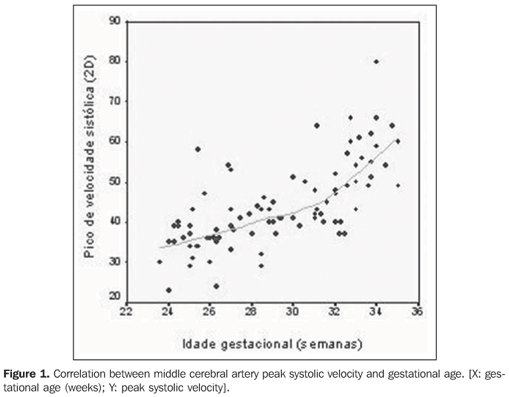
RESULTS The pregnancies were followed-up to the delivery. The mean gestational age at birth was 37 weeks and 2 days (28 weeks and 4 days to 41 weeks), and mean birth weight 2,877 g (1,400 g to 4,100 g). Mean hemoglobin concentration in newborns was 12.48g/dl (10.1 to 16.1 g/dl) and mean hematocrit level was 48.94% (37.1% to 62.0%). None of the newborns required transfusion at nursery and there were no stillborns or neonatal deaths. A strong correlation was observed between gestational age and MCA-PSV (r = 0.70; p < 0.001). The scatter plot between MCA-PSV and gestational age showed that such relation is non linear. The graph suggests an adjustment of exponential or polynomial regression between such data (Figure 1). The equation that best describes the relation between MCA-PSV and gestational age suggests an adjustment of cubic regression: Expected PSV = 44.5998 – 0.0874(IG)2 +
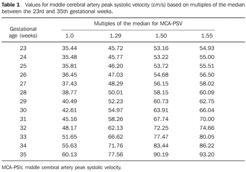
From this regression adjustment a table was built based on median multiples, according to the method adopted by Mari et al.(8). According to these authors, the median multiples are calculated by the ratio between the measured and expected values for a gestational age determined by the adjusted regression model. In this way, according to the results from a patient, the median multiple corresponds to a percentage of the expected value (values > 1 indicate that the observed result is higher than expected for that gestational age). Thus, the values of the "median" column correspond to the expected values and the others correspond to 29%, 50% and 55% above the expected value (Table 1).
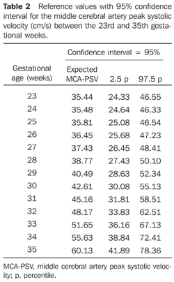
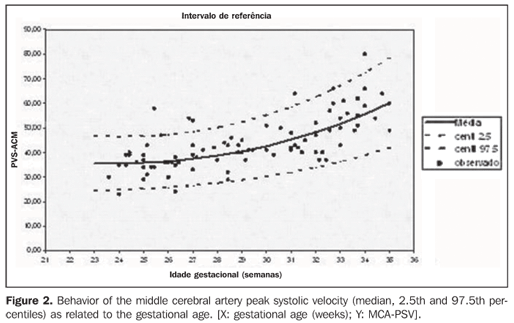
The determination of MCA-PSV reference values was based on the criteria proposed by Royston & Wright(9). The values are based on regression models for means and standard deviations (SD). A quadratic model was utilized for SD adjustment: [SD = 43.1872 – 2.8425(IG) + From the adjusted mean and SD for each studied gestational age, the percentiles were calculated according to the formula: Percentile = mean + K(SD) where: K is the percentile corresponding to the normal standard distribution. Table 2 demonstrates the 2.5th and 97.5th percentiles calculation, with a 95% confidence interval, using K = 1.96. Figure 2 demonstrates the behavior of the mean and of the 2.5th and 97.5th percentiles of MCA-PSV in relation to gestational age.
DISCUSSION Red blood cells alloimmunization remains a severe complication of pregnancy, in spite of the fact that its physiopathology has been recognized and its treatment, established. In developing countries, the incidence of such disorder has decreased very slowly, mainly due to insufficient prenatal prophylaxis with anti-D gammaglobulin. A previous study developed by the authors have found 39% premature births, 31% low birth weight, and 10% of newborns requiring blood transfusions in relation to a control group(10). Therefore the early detection of fetal hemolytic anemia, with prompt intervention by means of intrauterine transfusion or labor induction for neonatal therapy, may actually bring about good neonatal outcomes(11). The current invasive methods to quantify the degree of fetal anemia are the amniocentesis and cordocentesis. Amniocentesis presents questionable results when carried out before the 27th week(12); furthermore, this method may cause complications such as fetal-maternal hemorrhage, that may worsen the severity of the disease(13), and the need for serial amniocentesis(14). Cordocentesis presents a higher risk for fetal loss as compared with amniocentesis, and transplacental puncture is associated with fetal-maternal hemorrhage and increased sensitization(15). Noninvasive methods to measure the degree of fetal anemia present, as a greater benefit, the absence of maternal-fetal risks from the invasive methods. In the late eighties, several authors evaluated different fetal vessels with the Doppler mode in an attempt to develop a noninvasive method to quantify the fetal anemia degree in cases of red blood cells alloimmunization, but a consistent correlation between these parameters could not be find(5,16). In 1995 , Mari et al.(7) demonstrated increased blood flow velocity in anemic fetuses as compared with normal fetuses. For this measurement, these authors utilized the middle cerebral artery, as this vessel can be easily identified and its flow can be studied with a low and different from 0º angle. The MCAPSV can be accurately measured, with low intra and inter-observer variability. Later, several studies proved the superiority of MCA-PSV measurement over spectrophotometry in the prediction of fetal anemia(11,17–20). A study developed by the authors' group have evidenced higher hematocrit levels and lower need for blood transfusion in newborns from Rh alloimmunized pregnant women followed-up with MCA-PSV measurement than in those from mothers followed up with amniocentesis(21). In the present study, a strong correlation was observed between MCA-PSV and gestational age, with the best model being a exponential one. Such exponential model is in agreement with the reference curve of 135 pregnant women in the pioneer study developed by Mari et al.(7). This same exponential model has been obtained by Alshimmiri et al.(22) with 300 normal fetuses. However, in the study developed by Gadelha da Costa et al.(23), the authors obtained a linear relation between MCA-PSV and gestational age: MCA-PSV: 21.47 + 2.16GA. One objective of the present study was to determine a reference curve for MCA-PSV by using median multiples as proposed by Mari et al.(8). The authors determined values for the following median multiples: 1.0; 1.29; 1.50; 1.55. The value of 1.29 median multiple represents the cut-off for detection of mild anemia, 1.50 is the cut-off for the detection of moderate anemia and 1.55 is the cut-off for the detection of severe anemia, as proposed by Mari et al.(8). The authors have obtained 100% sensitivity, with 12% false-positive rate increased MCA-PSV in the prediction of moderate or severe fetal anemia. The comparison between the reference curves determined by the present study and by Mari et al.(8), demonstrated higher values for all gestational ages in the present study. Amongst the reasons for such differences, ethnical differences between both populations, nutritional levels of the patients, and the altitude of the places where the researches were developed, can be mentioned as factors that would justify the determination of a normality curve for different populations. Another objective of this study was the determination the reference values in percentiles for MCAPSV. The authors observed a progressive increase in MCA-PSV in the second half of pregnancies, in the same manner as observed by Gadelha da Costa et al.(23), Kurmanavicius et al.(24) and Bahlmann et al.(25). In a comparison with the study developed by Gadelha da Costa et al.(23) who have also evaluated a sample of the Brazilian population, the authors of the present study similar values for MCA-PSV in the period between the 22nd and 34th weeks. In comparison with the study developed by Kurmanavicius et al.(24) who have also adopted Royston & Wright(9) criteria for determination of the percentiles for MCA-PSV, the authors observed similar values in the studied gestational period, however, those authors found a linear correlation between MCAPSV values and gestational age, while in the present study a cubic equation was obtained. When comparing with the study of Bahlmann et al.(25), the values for MCA-PSV in the present study were higher for a same gestational period (23rd to 35th weeks). A possible explanation might be the method applied in that study, which utilized a 90% confidence interval, while in the present study, a 95% confidence interval was utilized. In summary, it is believed that the present study is relevant, considering that it is the second study determining a reference curve for MCA-PSV utilizing median multiples. The fact that this pilot study demonstrates different values as compared with those found by Mari et al.(8) justifies an ampler study involving the Brazilian population, preferably with a multi-centric approach to find out if these differences are due to ethnical factors. In anyway, the present study represented a first step towards the determination of a reference curve for MCA-PSV using median multiples, and that in the future it may prove useful in the follow-up of pregnant women with red blood cells alloimmunization.
REFERENCES 1. Liley AW. Liquor amnii analysis in the management of the pregnancy complicated by rhesus sensitization. Am J Obstet Gynecol. 1961;82:1359–70. [ ] 2. Simpson JL. Incidence and timing of pregnancy losses: relevance to evaluating safety of early prenatal diagnosis. Am J Med Genet. 1990;35:165–73. [ ] 3. MacKenzie IZ, Bowell PJ, Castle BM, et al. Serial fetal blood sampling for the management of pregnancies complicated by severe rhesus (D) isoimmunization. Br J Obstet Gynaecol. 1988;95:753–8. [ ] 4. Nicolaides KH, Soothill PW, Clewell WH, et al. Fetal haemoglobin measurement in the assessment of red cell isoimmunisation. Lancet. 1988;1:1073–5. [ ] 5. Nicolaides KH, Bilardo CM, Campbell S. Prediction of fetal anemia by measurement of the mean blood velocity in the fetal aorta. Am J Obstet Gynecol. 1990;162:209–12. [ ] 6. Copel JA, Grannum PA, Green JJ, et al. Pulsed Doppler flow-velocity waveforms in the prediction of fetal hematocrit of the severely isoimmunized pregnancy. Am J Obstet Gynecol. 1989; 161:341–4. [ ] 7. Mari G, Adrignolo A, Abuhamad AZ, et al. Diagnosis of fetal anemia with Doppler ultrasound in the pregnancy complicated by maternal blood group immunization. Ultrasound Obstet Gynecol. 1995;5:400–5. [ ] 8. Mari G, Deter RL, Carpenter RL, et al. Noninvasive diagnosis by Doppler ultrasonography of fetal anemia due to maternal red-cell alloimmunization. N Engl J Med. 2000;342:9–14. [ ] 9. Royston P, Wright EM. How to construct 'normal ranges' for fetal variables. Ultrasound Obstet Gynecol. 1998;11:30–8. [ ] 10. Nardozza LM, Camano L, Moron AF, et al. Pregnancy outcome for Rh-alloimmunized women. Int J Gynaecol Obstet. 2005;90:103–6. [ ] 11. Oepkes D. Invasive versus non-invasive testing in red-cell alloimunized pregnancies. Eur J Obstet Gynecol Reprod Biol. 2000;92:83–9. [ ] 12. Rahman F, Detti L, Ozcan T, et al. Can a single measurement of amniotic fluid delta optical density be safely used in the clinical management of Rhesus-alloimmunized pregnancies before 27 weeks' gestation? Acta Obstet Gynecol Scand. 1998;77:804–7. [ ] 13. Bowman JM, Pollock JM. Transplacental fetal hemorrhage after amniocentesis. Obstet Gynecol. 1985;66:749–54. [ ] 14. Queenan JT, Tomai TP, Ural SH, et al. Deviation in amniotic fluid optical density at a wavelength of 450 nm in Rh-immunized pregnancies from 14 to 40 weeks' gestation: a proposal for clinical management. Am J Obstet Gynecol. 1993;168:1370–6. [ ] 15. MacGregor SN, Silver RK, Sholl JS. Enhanced sensitization after cordocentesis in a rhesus-isoimmunized pregnancy. Am J Obstet Gynecol. 1991;165:382–3. [ ] 16. Rightmire DA, Nicolaides KH, Rodeck CH, et al. Fetal blood velocities in Rh isoimmunization: relationship to gestational age and to fetal hematocrit. Obstet Gynecol. 1986;68:233–6. [ ] 17. Nishie EN, Brizot ML, Liao AW, et al. A comparison between middle cerebral artery peak systolic velocity and amniotic fluid optical density at 450 nm in the prediction of fetal anemia. Am J Obstet Gynecol. 2003;188:214–9. [ ] 18. Oepkes D, Seaward PG, Vandenbussche FP, et al. Doppler ultrasonography versus amniocentesis to predict fetal anemia. N Engl Med J. 2006;355:156–64. [ ] 19. Pastore AR. Dopplervelocimetria da artéria cerebral média fetal: o divisor de águas no diagnóstico da anemia fetal. Radiol Bras. 2006;39(1):iii. [ ] 20. Nardozza MM, Camano L, Moron AF, et al. Alterações ultra-sonográficas na gravidez Rh negativo sensibilizada avaliada pela espectrofotometria do líquido amniótico e pela dopplervelocimetria da artéria cerebral média. Radiol Bras. 2006; 39:11–3. [ ] 21. Nardozza LM, Moron AF, Araujo Júnior E, et al. Rh alloimmunization: Doppler or amniotic fluid analysis in the prediction of fetal anemia? Arch Gynecol Obstet. 2007;275:107–11. [ ] 22. Alshimmiri MM, Hamoud MS, Al-Saleh EA, et al. Prediction of fetal anemia by middle cerebral artery peak systolic velocity in pregnancies complicated by Rhesus isoimmunization. J Perinatol. 2003;23:536–40. [ ] 23. Gadelha Da Costa A, Mauad Filho F, Spara P, et al. Fetal hemodynamics evaluated by Doppler velocimetry in the second half of pregnancy. Ultrasound Med Biol. 2005;31:1023–30. [ ] 24. Kurmanavicius J, Streicher A, Wright EM, et al. Reference values of fetal peak systolic blood flow velocity in the middle cerebral artery at 19–40 weeks of gestation. Ultrasound Obstet Gynecol. 2001;17:50–3. [ ] 25. Bahlmann F, Reinhard I, Krummenauer F, et al. Blood flow velocity waveforms of the fetal middle cerebral artery in a normal population: reference values from 18 weeks to 42 weeks of gestation. J Perinat Med. 2002;30:490–501. [ ] Received March 18, 2008. Accepted after revision May 30, 2008. * Study developed in the Division of Fetal Medicine of Department of Obstetrics at Universidade Federal de São Paulo/Escola Paulista de Medicina (Unifesp/EPM), São Paulo, SP, Brazil. |
|
Av. Paulista, 37 - 7° andar - Conj. 71 - CEP 01311-902 - São Paulo - SP - Brazil - Phone: (11) 3372-4544 - Fax: (11) 3372-4554


