Radiologia Brasileira - Publicação Científica Oficial do Colégio Brasileiro de Radiologia
AMB - Associação Médica Brasileira CNA - Comissão Nacional de Acreditação
 Vol. 41 nº 6 - Nov. / Dec. of 2008
|
ORIGINAL ARTICLE
|
|
The role of magnetic resonance cholangiography in the evaluation of biliary anatomy in living liver donors |
|
|
Autho(rs): Elaine Cristina de Moraes Arruda, Julio Cezar Uili Coelho, Jorge Massayuki Yokochi, Jorge Eduardo Fouto Matias |
|
|
Keywords: Liver transplant, Living donors, Magnetic resonance imaging, Biliary tract |
|
|
Abstract:
IMaster, MD, Pediatric Radiologist at Instituto de Radiodiagnóstico – Ultra-X, São José do Rio Preto, SP, Brazil
INTRODUCTION Liver transplant represents the sole treatment possibility for many patients affected by irreversible hepatic diseases, but the scarce offering of organs has been one of the limiting factors in the survival of waiting-list patients with hepatic failure(1). Living-donor liver transplant is a definite method of treatment introduced for minimizing this problem(2). Particular care should be dedicated to the planning for management of the biliary ducts during liver lobe resection and implantation, considering that the high variability of the biliary anatomy in the population and the pattern of second order biliary branches could change the surgical technique or even represent a contraindication for liver donation(3). Anatomic variants have been described in as many as 19.7%(4) to 43%(5) of individuals. Studies have evidenced the utilization of magnetic resonance imaging as the method of choice for preoperative evaluation of the biliary anatomy(2,6–8) and also as a sole method for preoperative evaluation in these cases, considering its accuracy in the detection of hepatic parenchyma abnormalities hepatic and lobar volume, besides the depiction of the portal artery, and of the venous and biliary systems anatomy(9–11). However, the utilization of this method for this purpose still remains to be more deeply evaluated. The present study was aimed at retrospectively evaluating the accuracy of magnetic resonance cholangiography (MR cholangiography) for depicting the biliary tract in living liver donors in correlation with intraoperative findings.
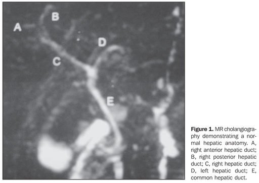
MATERIALS AND METHODS MR cholangiography images and records of all living liver donors were reviewed in the period between November 1998 and May 2006. The present casuistic included 50 donors. All the living donors for liver transplant were included. The individuals with significant vascular or biliary anomalies resulting in contraindication for liver donation were excluded because of the increased risk for postoperative complications. Also, one donor whose records had not been found was excluded. The present study was approved by the Committee for Ethics in Research in Humans of the Institution (Register No. CEP/HC 759.178/2003-11). The studies were performed in a Gyroscan ACS15 model system (Philips Medical Systems; Best, The Nederlands), with 1.5 T magnetic field and body coil. The examination protocol included the following sequences: – Axial, coronal and sagittal, turbo field echo, T1-weighted sequences, adopting the following parameters: 11/4 msec repetition time (TR)/echo time (TE); flip angle = 25°. – Axial turbo spin eco (TSE) T2-weighted sequence for evaluating the liver (TR/TE: 1,800/160 msec; thickness: 8 mm; gap: 0.8; matrix: 258 × 205 reconstructed for 512 × 410; field of view (FOV): ranging between 300 mm and 380 mm, according to the patient; number of signal averaged (NSA): 4; number of sections: 24). – Coronal, with overcontinuous slices, 1.5 mm overlapping, with inversion recovery technique for fat suppression, T2-weighted STIR (TSE) for evaluating the biliary tract (TR/TE: 1,800/500 msec; FOV: 230 mm; inversion time: 160 msec; matrix: 256 × 179; NSA: 2; thickness: 3 mm; number of sections: 65 to 80). The sequences were acquired with respiratory gating mode, and the total examination time ranged between 30 and 50 minutes, depending on the patient´s respiration regularity. Immediately after acquisition, the images were reconstructed and transmitted to the console for processing. The biliary tract anatomy was reviewed on the images acquired and based on maximum-intensity-projection reconstruction. The images were reviewed both in workstations and hardcopies. All the images were reviewed by a same specialized radiologist, with about ten years experience in the interpretation of abdominal images. The analysis included an evaluation of any variations, particularly those involving the presence of segmental ducts of the right and left lobes, or even the presence of common hepatic duct trifurcation, and the evaluation of the choledocal duct and its main branches. The extrahepatic biliary tract was considered as normal in the presence of only one right hepatic duct and one left hepatic duct joining to form the common hepatic duct, and the cystic duct joining the common hepatic duct at right (Figure 1). The intraoperative findings described by the surgeon were considered as a reference pattern. Only those anatomical alterations that affected the surgical strategy and had not been previously observed at magnetic resonance cholangiography were considered as being in disagreement.
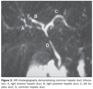
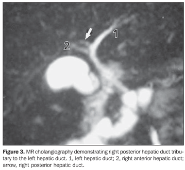
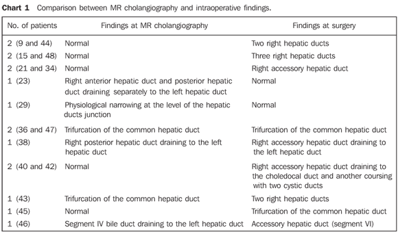
RESULTS Six of the 50 cases involved pediatric liver transplants (recipients with < 18 years of age) and 44, adult liver transplants. The donors ranged in age from 18 to 60 years (mean = 32.4 years). Thirty-one patients (62%) were male, with mean age of 30.8 years. Female patients were 19 (38%), with mean age of 35.2 years. Three types of grafts were utilized as follows: Couinaud liver segments II and III (lateral left segmentectomy) in two cases; Couinaud segments II, III and IV (left lobectomy) in one case; and Couinaud segments V, VI, VII and VIII (right lobectomy) in 47 cases. Anatomical alterations which affected the surgical strategy were found in only two right-lobe donors. Normal hepatic anatomy could be found by MR cholangiography in 43 patients, and anatomical alterations were found in seven (14%). Three donors (6%) presented junction of the right anterior and posterior hepatic ducts with the left hepatic duct (common hepatic duct trifurcation) (Figure 2). In one case (2%), the right posterior hepatic duct was tributary of the left hepatic duct (Figure 3). In one case (2%) the right posterior and anterior hepatic ducts drained separately into the left hepatic duct. In one case (2%) physiological narrowing at the level of the hepatic ducts junction. Finally, in one case (2%) the bile duct of the segment IV drained to the left hepatic segment, near the junction with the right hepatic duct.
Intraoperative findings corresponding to biliary tract alterations were described in 14 donors (28%). Three patients (6%) presented common hepatic duct trifurcation; three (6%) presented right hepatic duct duplication; and two patients (4%), right hepatic triplication, one of these patients with also an accessory duct. In one donor (2%), a fine accessory bile duct originating from the right posterior duct was observed. Five donors (10%) presented an accessory right hepatic duct, one of them with the accessory duct tributary of the left hepatic duct, another with the accessory duct draining into the choledocal duct, and another with two cystic ducts besides the accessory hepatic duct. In the analysis of the 50 donors, 34 presented normal anatomy at the surgery, in agreement with the findings at MR cholangiography. Anatomical variations were found at MR cholangiography in seven donors (14%), and in 14 donors (28%) during surgery. These variations were found in two cases with right hepatic ducts, three with triplication of right hepatic duct, one case of common hepatic duct trifurcation, and four cases with accessory hepatic duct. In one of these donors, the accessory duct was tributary of the choledocal duct, in another, of the left hepatic duct, and another with the presence of two cystic ducts. In one donor who had a very small accessory duct that was linked during the surgery, this finding was not considered as being in disagreement. In two patients (4%) anatomical variations were found at MR cholangiography, but intraoperative findings demonstrated a normal anatomy. In one donor, the right anterior and posterior ducts drained separately into the left hepatic duct, and in another case, non relevant from the surgical point of view, a physiological narrowing was described at the level of the hepatic ducts junction (also this case was not considered as being in disagreement). Among the seven studies with biliary tract abnormalities, five presented results in agreement with the intraoperative findings, two of them with common hepatic duct trifurcation, and one with the right posterior hepatic ducts draining into the left hepatic ducts (the latter demonstrating an accessory duct draining to the left hepatic duct at surgery, interpreted as being in agreement, considering its non-relevance from the surgical point of view). In two donors, the MR cholangiography demonstrated anatomic alterations which although not properly corresponding to the intraoperative findings, neither resulted in change of the surgical strategy nor in the planned anastomotic changes. In one of these donors, MR cholangiography demonstrated trifurcation, but two right hepatic ducts were present, and in the other with an accessory hepatic duct, it was interpreted as a biliary duct of the segment IV. The comparison between MR cholangiography and intraoperative findings is shown on Chart 1.
Therefore, 41/50 donors (82%) presented correspondence between imaging and intraoperative findings, and 9/50 (18%) did not. The MR cholangiography sensitivity was 43%, specificity, 97%, positive predictive value, 86%, negative predictive value, 81%, and accuracy, 81.6%.
DISCUSSION Living-donor liver transplant is a definite method of treatment for patients with irreversible hepatic diseases, particularly in countries where cadaver liver donors are scarce or even non-existent(12). In Brazil, the number of living liver donors increased 10% in 2005, and in 2006 remained stable(13). The safety of the donors is extremely important, considering that they are healthy individuals submitted to an extensive surgical procedure. So, the detection of the biliary tract anatomy is crucial for allowing the surgical planning and avoiding unnecessary surgery in donors with anatomical variations, besides preventing possible postoperative complications both for liver donors and recipients(7). According to Liu et al.(14), biliary complications remain as the most noticeable weakness in living-donor liver transplant, playing a significant role in the occurrence of postoperative morbidities occasionally caused by graft loss. According to Marcos et al.(15), biliary reconstruction corresponds to the most challenging part of the surgery in the liver recipient, considering that double or triple anastomosis certainly represents a risk factor for biliary complications(16). Biliary anatomical variations which lack pathological meaning in the general population, assume a greater relevance in cases of right lobe donation. These variations include common hepatic duct trifurcation, accessory right hepatic duct and drainage from the right anterior or posterior segmental duct directly into the right hepatic duct. Although such variations do not contraindicate liver donation, the preoperative identification prevents that these ducts are inadvertently connected, resulting in atrophy of the involved portions of the liver(6). Retrograde endoscopic cholangiography or percutaneous transhepatic cholangiography are considered as "golden-standard" methods for evaluating the biliary tract, but are not routinely performed because of their invasiveness and association with high risks for complications(8,14,17). The rate of complications from retrograde endoscopic cholangiography ranges between 0.5% and 5%, and from percutaneous transhepatic cholangiography, 3.4%(17). Recently, some studies reported magnetic resonance imaging as the sole preoperative method for evaluating living liver donor candidates, demonstrating good results in the evaluation of the biliary tract(9,11,18). This assumption is based on the fact that conventional MR cholangiography T2weighted sequences demonstrate high signal intensity from static fluid structures while the background signal is suppressed(19). On the other hand, there is a difficulty in the evaluation of a non-dilated biliary tract(3,19). Innovations such as the utilization of a biliary contrast agent (mangafodipir trisodium) have allowed the acquisition of higher resolution images, with good results because of the better enhancement of the biliary tract and higher differentiation from the hepatic parenchyma and from the vascular system(7,8,20,21). Ayuso et al. have observed a sensitivity of 93.7% and specificity of 100%(22). However, high cost, limited contrast agent availability, possibility of allergic reactions and increased images acquisition time constitute limiting factor for the utilization of this method(21). Other authors have compared contrast-enhanced (gadobenate dimeglumine – Gd-DTPA) MR cholangiography T1-weighted sequences with findings at conventional MR cholangiography T2-weighted sequences and there was a preponderant preference for contrast-enhanced MR cholangiography in the evaluation of the biliary tract(23,24). This contrast agent combines the properties of a gadolinium-based extracellular contrast agent as a hepatocyte-direct excreted at about 2%–4% through the biliary tract. An et al(25) have described an accuracy of 75% with MR cholangiography T2-weighted sequences, 79% with paramagnetic contrast-enhanced T1 weighted sequences, and an increase to 92% in accuracy with the evaluation by means of a combination of both methods. The results of the present study, likewise those previously reported by Lee et al.(9) reflect a diligent analysis in relation to the consistency of MR cholangiography for appropriately visualizing the biliary anatomy in liver donors. The present study could demonstrate that MR cholangiography presents a good reproducibility in relation to the surgical findings. However, the low sensitivity of the method and the failure in detecting anatomical variations in nine cases (18%), among them, the presence of common hepatic duct trifurcation, duplicated or triplicated right hepatic ducts and accessory hepatic ducts, inspires prudence and demonstrates that the segmental ducts definition is not clear. As previously reported, the utilization of MR cholangiography with specific contrast agents for studying the biliary tract, as well as further investigation about the utilization of paramagnetic contras agents such as gadobenate dimeglumine have contributed for improvement of the method. Recently, single-shot fast spin echo sequences were adopted as a standard method for MR cholangiography(26), and new sequences such as half Fourier RARE have demonstrated technical advances, allowing imaging of the whole biliary tract during a single 18-second breath-hold(27). Maybe the utilization of the described sequences, if available, could result in better images as those already reported by some authors(9,11). Further studies are required to evaluate technological developments such as new sequences and utilization of paramagnetic and biliary contrast agents. Considering the retrospective character o the present study, the authors could not determine the number of patients excluded as well as the identified anatomical variations. It can be concluded that MR cholangiography performed with T2-weighted sequences presented a high accuracy, high specificity and low sensitivity. MRI is a safe and non-invasive method with potential capacity and applicability in the preoperative evaluation of the biliary tract in living liver donors. Continued technical innovations will certainly allow an expansion of the utilization of this method in a near future.
REFERENCES 1. Emond JC, Renz JF, Ferrel LD, et al. Functional analysis of grafts from living donors. Ann Surg. 1996;224:544–54. [ ] 2. Bassignani MJ, Fulcher AS, Szucs RA, et al. Use of imaging for living donor liver transplantation. Radiographics. 2001;21:39–52. [ ] 3. Yeh BM, Breiman RS, Taouli B, et al. Biliary tract depiction in living potential liver donors: comparison of conventional MR, mangafodipir trisodium-enhanced excretory MR, and multi-detector row CT cholangiography – initial experience. Radiology. 2004;230:645–51. [ ] 4. Chisuwa H, Hashikura Y, Mita A, et al. Living liver donation: preoperative assessment, anatomic considerations, and long-term outcome. Transplantation. 2003;75:1670–6. [ ] 5. Couinaud C. Le foie: études anatomiques et chirurgicales. Paris: Masson; 1957. [ ] 6. Fulcher AS, Szucs RA, Bassignani MJ, et al. Right lobe living donor liver transplantation: preoperative evaluation of the donor with MR imaging. AJR Am J Roentgenol. 2001;176:1483–91. [ ] 7. Kapoor V, Peterson MS, Baron RL, et al. Intrahepatic biliary anatomy of living adult liver donors: correlation of mangafodipir trisodium-enhanced MR cholangiography and intraoperative cholangiography. AJR Am J Roentgenol. 2002; 179:1281–6. [ ] 8. Kim RD, Sakamoto S, Haider MA, et al. Role of magnetic resonance cholangiography in assessing biliary anatomy in right lobe living donors. Transplantation. 2005;79:1417–21. [ ] 9. Lee VS, Morgan GR, Teperman LW, et al. MR imaging as the sole preoperative imaging modality for right hepatectomy: a prospective study of living adult-to-adult liver donor candidates. AJR Am J Roentgenol. 2001;176:1475–82. [ ] 10. Goyen M, Barkhausen J, Debatin JF, et al. Right-lobe living related liver transplantation: evaluation of a comprehensive magnetic resonance imaging protocol for assessing potential donors. Liver Transpl. 2002;8:241–50. [ ] 11. Cheng YF, Chen CL, Huang TL, et al. Single imaging modality evaluation of living donors in liver transplantation: magnetic resonance imaging. Transplantation. 2001;72:1527–33. [ ] 12. Hashikura Y, Makuuchi M, Kawasaki S, et al. Successful living-related partial liver transplantation to an adult patient. Lancet. 1994;343:1233–4. [ ] 13. Associação Brasileira de Transplante de Órgãos. Área para profissionais. Gráficos 2005/2006. [acessado em: 9/10/2007]. Disponível em: http://www.abto.org.br/profissionais.asp [ ] 14. Liu CL, Lo CM, Chan SC, et al. The right may not be always right: biliary anatomy contraindicates right lobe live donor liver transplantation. Liver Transpl. 2004;10:811–2. [ ] 15. Marcos A, Ham JM, Fisher RA, et al. Surgical management of anatomical variation of the right lobe in living donor liver transplantation. Ann Surg. 2000;231:824–31. [ ] 16. Fan ST, Lo CM, Liu CL, et al. Biliary reconstruction and complications of right lobe live donor liver transplantation. Ann Surg. 2002;236:676–83. [ ] 17. Caoili EM, Paulson EK, Heyneman LE, et al. Helical CT cholangiography with three-dimensional volume rendering using an oral biliary contrast agent: feasibility of a novel technique. AJR Am J Roentgenol. 2000;174:487–92. [ ] 18. Sahani D, D'souza R, Kadavigere R, et al. Evaluation of living liver transplant donors: method for precise anatomic defining by using a dedicated contrast-enhanced MR imaging protocol. Radiographics. 2004;24:957–67. [ ] 19. Song GW, Lee SG, Hwang S, et al. Preoperative evaluation of biliary anatomy of donor in living donor liver transplantation by conventional nonenhanced magnetic resonance cholangiography. Transpl Int. 2007;20:167–73. [ ] 20. Lee VS, Rofsky NM, Morgan GR, et al. Volumetric mangafodipir trisodium-enhanced cholangiography to define intrahepatic biliary anatomy. AJR Am J Roentgenol. 2001;176:906–8. [ ] 21. Lee VS, Krinsky GA, Nazarro CA, et al. Defining intrahepatic biliary anatomy in living liver transplant donor candidates at mangafodipir trissodium-enhanced MR cholangiography versus conventional T2-weighted MR cholangiography. Radiology. 2004;233:659–66. [ ] 22. Ayuso JR, Ayuso C, Bombuy E, et al. Preoperative evaluation of biliary anatomy in adult live liver donors with volumetric mangafodipir trisodium enhanced magnetic resonance cholangiography. Liver Transpl. 2004;10:1391–7. [ ] 23. Lim JS, Kim MJ, Kim JH, et al. Preoperative MRI of potential living-donor-related liver transplantation using a single dose of gadobenate dimeglumine. AJR Am J Roentgenol. 2005;185: 424–31. [ ] 24. Papanikolaou N, Prassopoulos P, Eracleous E, et al. Contrast-enhanced magnetic resonance cholangiography versus heavily T2-weighted magnetic resonance cholangiography. Invest Radiol. 2001;36:682–6. [ ] 25. An, SK, Lee JM, Suh KS, et al. Gadobenate dimeglumine-enhanced liver MRI as the sole preoperative imaging technique: a prospective study of living liver donors. AJR Am J Roentgenol. 2006;187:1223–33. [ ] 26. Irie H, Honda H, Kuroiwa T, et al. Pitfalls in MR cholangiopancratographic interpretation. Radiographics. 2001;21:23–37. [ ] 27. Fulcher AS, Turner MA, Capps GW. MR cholangiography: technical advances and clinical applications. Radiographics. 1999;19:25–44. [ ] Received December 16, 2007. Accepted after revision May 27, 2008. * Study developed at Hospital de Clínicas da Universidade Federal do Paraná (UFPR), Curitiba, PR, Brazil. |
|

Av. Paulista, 37 - 7° andar - Conj. 71 - CEP 01311-902 - São Paulo - SP - Brazil - Phone: (11) 3372-4544 - Fax: (11) 3372-4554