Radiologia Brasileira - Publicação Científica Oficial do Colégio Brasileiro de Radiologia
AMB - Associação Médica Brasileira CNA - Comissão Nacional de Acreditação
 Vol. 41 nº 5 - Sep. / Oct. of 2008
|
ORIGINAL ARTICLE
|
|
Portal blood flow volume measurement in schistosomal patients: evaluation of Doppler ultrasonography reproducibility |
|
|
Autho(rs): Alberto Ribeiro de Souza Leão, José Eduardo Mourão Santos, Danilo Sales Moulin, David Carlos Shigueoka, Ramiro Colleoni, Giuseppe D'Ippolito |
|
|
Keywords: Portal flow, Measurement, Doppler ultrasonography, Reproducibility, Portal hypertension |
|
|
Abstract:
IMDs, Residents in Radiology, Fellows PhD degree, Department of Imaging Diagnosis at Universidade Federal de São Paulo/Escola Paulista de Medicina (Unifesp/EPM), São Paulo, SP, Brazil
INTRODUCTION In healthy patients, the porto-hepatic circulation compensates great variations in the blood flow with small variations in the portal pressure(1). Pathological alterations in the hemodynamics of this system are characterized by a chronic increase in the portal venous pressure defined as portal hypertension secondary to an interference with the splanchnic venous blood flow and clinically translated into collateral circulation visible on the abdominal wall, ascites and esophagogastric alterations, i.e., esophageal varices, gastric varices and congestive gastropathy(1). Cirrhosis, the hepatosplenic form of schistosomiasis mansoni, liver, biliary tract or pancreas neoplasms, portal vein thromboembolic phenomena and suprahepatic diseases such as right heart failure and inferior vena cava occlusion by thrombi or tumors are particularly important amongst diseases that may course with portal hypertension(2). The diagnosis of portal hypertension can be noninvasively performed through correlation between semiological data and complementary methods, as well as invasively, either by direct approach with surgical measurement of the portal venous pressure or by indirect approach with the measurement of the occluded and free hepatic venous pressure aiming at obtaining the hepatic venous pressure gradient(3) corresponding to the difference between both pressures, that in healthy individuals should be < 5 mmHg(4). The direct measurement of the portal venous pressure is the most effective method for evaluating its actual elevation. The measurement of the portal venous pressure levels can aid in the differential diagnosis of the causes for portal hypertension, in the evaluation of the risk for bleeding due to gastroesophageal varices rupture that is the main cause for morbimortality in these patients, in the evaluation of the medicamentous therapy effectiveness, in the prophylaxis of gastroesophageal varices bleeding, in the therapeutic decision making in cases of liver resection, and in the evaluation o the patients' prognosis(3,5). Notwithstanding the unquestionable applications of the hepatic venous pressure gradient, high cost and operator dependence have limited the availability of this method. Additionally, despite its lower aggressiveness as compared with the other techniques for direct measurement of the portal venous pressure, this method is still invasive. For these reasons, the identification of a noninvasive marker for portal hypertension still remains as a challenge. Several authors have suggested that some Doppler ultrasound (US) parameters could have a prognostic value and usefulness in the evaluation of the risk for bleeding of esophageal varices. However, this technique has been sub utilized for this purpose and its clinical utility has been debated(3). Amongst hemodynamic parameters measurable at Doppler US, peak portal flow velocity is the most frequently adopted, but its utilization is limited by the overlap between pathological and physiological values(6). Additionally, studies utilizing pressure gradient monitoring have demonstrated the absence of a direct relationship between variations in flow velocity and pressoric levels(7). The noninvasive measurement of the portal vein flow volume in patients with portal hypertension has gained ground as an alternative method for diagnostic anamnesis and follow-up of these patients. Studies evaluating cirrhotic patients have demonstrated a significant influence of the portal venous flow on the functional activity of the liver(5). The intraoperative measurement of the portal venous flow in pediatric patients submitted to liver transplantation can predict which of them are likely to develop portal thrombosis(8). Additionally, results of preliminary studies suggest that, among several variables, portal flow volume seems to be a good marker for hepatic function(9). On the other hand, schistosomal portal hypertension has been considered as an ideal model for the study of portal flow physiology(5). Studies in the literature focused on different aspects of the disease have already evaluated the reproducibility of noninvasive methods for evaluation of schistosomal patients. Magnetic resonance imaging (MRI) reproducibility in the evaluation of morphological alterations of the liver and spleen in these patients has been evaluated with high levels of agreement(10), demonstrating high reliability in the quantification of the portal flow in healthy patients, with higher interobserver agreement as compared with Doppler US(11). Intermethod agreement was low in the portal flow quantification(11). The same levels of agreement were observed in the evaluation of periportal fibrosis(12,13). Despite the wide availability and widespread use of the method, the literature demonstrates controversial results in relation to the Doppler US reproducibility in the measurement of several dopplerfluxometric parameters, among them the flow volume, utilized in the evaluation of the portal vein(9,14). The present study is aimed at evaluating Doppler US reproducibility in the measurement of the portal flow in patients with schistosomal portal hypertension. The option for this model was based on the high incidence of portal hypertension, considering that schistosomiasis still remains as the main cause for this complication world-wide(5), with no other similar study being found in the literature.
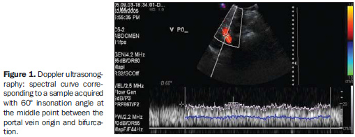
MATERIALS AND METHODS A prospective, transversal, observational, double-blinded and self-paired study was developed with 21 patients referred by the ambulatory of schistosomiasis of the Division of Gastroenterology - Universidade Federal de São Paulo/Escola Paulista de Medicina (Unifesp/EPM), and submitted to US scans in the period between February 2005 and December 2006. The study was previously approved by the Committee for Ethics in Research of the institution and all the patients signed a term of free and informed consent. The diagnosis of schistosomiasis mansoni was based on the results of rectal biopsy or a strong clinical/laboratory evidence (signs of portal hypertension and/or positive proctoparasitological stool tests) and epidemiological evidences (contact with water of ponds and rivers in endemic areas). The patients were evaluated after 6 to 8-hour fasting by Doppler US. Scanning and interpretation of images were performed and analyzed by three independent observers (observers 1, 2 and 3), all of them with at least three years of experience in abdominal Doppler US and having accomplished medical residency in imaging diagnosis with specific training for portal flow measurement. The Doppler US studies were performed in an EnVisorTM equipment (Philips Medical Systems; Bothell, USA), utilizing a multi frequency convex array abdominal transducer, following the standard imaging views recommended by the World Health Organization for sonographic evaluation of the liver, spleen and splanchnic vascular system. The three readings were sequentially performed with a 10-minute interval (time required for changing from one observer to another). Doppler US examinations of the portal vein were performed with the patients in dorsal decubitus, after a short rest period, with oblique subcostal and intercostal views of the portal vein trunk at a middle distance from the bifurcation, within a same respiratory phase, and with an insonation angle ranging between 45-60º (Figure 1), and 20-30-minute acquisition time. The inner diameter and the cross-section area of the portal vein were measured on the same topography utilized for dopplerfluxometric analysis. The spectral interval selected for analysis was of at least four seconds. Once the parameters required by the equipment software were provided, the values of the flow volume for the respective sample were obtained.
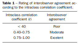
Statistical analysis Tests for numeric variables were utilized for evaluating the agreement among the different readings. For data analysis the authors utilized the intraclass coefficient calculation based on a 95% confidence interval (CI at 95 presented a p-value < 5% [p < 0.05]) and the t-paired test interpreted according the classification proposed by Fleiss (1981), as shown on Table 1. Also, the linearity degree between measurements obtained by two observers through the Pearson's linear correlation coefficient, considering 1 (r = 1) as the perfect positive correlation between two variables.
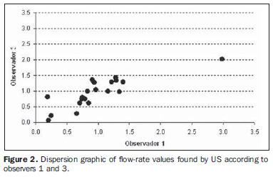
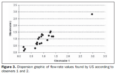
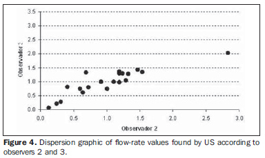
RESULTS The paired analysis of the reproducibility of Doppler US studies performed by the three observers demonstrated a good interobserver agreement. The intraclass correlation coefficient ranged between 80.6% and 93.0% (CI at 95% [65.3%; 95.8%]), with the Pearson's correlation coefficient ranging between 81.6% and 92.7% (Table 2). Based on the t-paired test, one may observe that generally, on average, the evaluation of US results demonstrated agreement, considering that the statistical test has not shown a significant difference in the mean portal flow evaluated by the observers (p = 0.954 / 0.758 / 0.749).
According to the dispersion graphics (Figures 2, 3 and 4), a high correlation was observed among measurements performed by the observers.
DISCUSSION The consolidation of alternative non-invasive methods for measuring the portal pressure for diagnosis purposes, as well as the endoscopic screening for gastroesophageal varices in the prophylaxis of the risk for high digestive bleeding, constitute a permanent objective for aiding in the management of patients with portal hypertension(15). Recent studies advocate that non-invasive methods have shown to be effective as a diagnostic alternative for measurement of the mean portal venous flow volume in patients with clinical suspicion of portal hypertension, for evaluation of the degree of impairment of the hepatic function and risk for high digestive bleeding, besides representing a measurable variable to be taken into consideration in the approach of patients submitted to liver transplantation, predicting the graft feasibility and the risk for portal venous thrombosis(5,8,9). Presently, with its wide availability and low cost, US has been considered as the non-invasive diagnostic method of choice for approaching patients with portal hypertension. Questions still remain, however, as to errors in the measurement of the area of cross-section of the vessel, interobserver variability, changes associated with physiological events and patient's biotype, besides the utilization of overlapping parameters such as velocity and flow rates(6,14,16-21). Considering that the diagnostic accuracy of a method is an essential parameter for determining its usefulness that may be defined by the evaluation of its reproducibility (interobserver agreement)(22), it is necessary to validate non-invasive methods (US, for example) capable of evaluating hemodynamic parameters in patients with portal hypertension. The present study was aimed at evaluating the Doppler US reproducibility in the measurement of portal venous flow volume. The option for patients with portal hypertension secondary to schistosomiasis was based on the wide range and variability of flow volume rates observed in these patients as a typical sign of their hemodynamic pattern of portal hyperflow, representing an ideal model for research and evaluation of this method accuracy(5). The evaluation of Doppler US reproducibility in the measurement of the mean portal venous flow volume in these patients demonstrated a good interobserver agreement which has not been previously described in the literature. Considering the wide availability and excellent cost/benefit ratio of the method, besides the high prevalence (in the case of schistosomiasis) of the disease in developing countries, evidences of a good reproducibility reinforce the role of a diagnostic method in the anamnesis of these patients. The present study presented some limitations such as the size of the sample (21 patients) and the observers' awareness of the patients' condition (schistosomiasis), besides the technical difficulties inherent in the morphological alterations of the hepatic and vascular systems typically found in advanced stages of this disease. Finally, the present study demonstrated a good reproducibility of Doppler US in the measurement of the portal venous flow volume in patients with portal hypertension secondary to schistosomiasis, adding value to this widely diffused method in the evaluation of this variable. Further studies establishing physiological and pathological values for portal venous flow may consolidate the role of Doppler US in the diagnostic and prognostic approach to alterations in the hemodynamics of this system.
REFERENCES 1. Bem RS, Lora FL, Souza RCA, et al. Correlação das características do ecodoppler do sistema porta com presença de alterações endoscópicas secundárias à hipertensão porta em pacientes com cirrose hepática. Arq Gastroenterol. 2006;43:178-83. [ ] 2. Petroianu A. Tratamento cirúrgico da hipertensão porta na esquistossomose mansoni. Rev Soc Bras Med Trop. 2003;36:253-65. [ ] 3. Dittrich S, Mattos AA, Cheinquer H, et al. Correlação entre a contagem de plaquetas no sangue e o gradiente de pressão venosa hepática em pacientes cirróticos. Arq Gastroenterol. 2005;42:35-40. [ ] 4. Dib N, Oberti F, Calès P. Current management of the complications of portal hypertension: variceal bleeding and ascites. CMAJ. 2006;174:1433-43. [ ] 5. Alves Jr A, Fontes DA, Melo VA, et al. Hipertensão portal esquistossomótica: influência do fluxo sangüíneo portal nos níveis séricos das enzimas hepáticas. Arq Gastroenterol. 2003;40:203-8. [ ] 6. Machado MM, Rosa ACF, Barros N, et al. Estudo Doppler na hipertensão portal. Radiol Bras. 2004;37:35-9. [ ] 7. Choi YJ, Baik SK, Park DH, et al. Comparison of Doppler ultrasonography and the hepatic venous pressure gradient in assessing portal hypertension in liver cirrhosis. J Gastroenterol Hepatol. 2003;18:424-9. [ ] 8. Bueno J, Escartín A, Balsells J, et al. Intraoperative flow measurement of native liver and allograft during orthotopic liver transplantation in children. Transplant Proc. 2007;39:2278-9. [ ] 9. Gontarczyk GW, Łagiewska B, Pacholczyk M, et al. Intraoperative blood flow measurements and liver allograft function: preliminary results. Transplant Proc. 2006;38:234-6. [ ] 10. Bezerra ASA, D'Ippolito G, Caldana RP, et al. Avaliação hepática e esplênica por ressonância magnética em pacientes portadores de esquistossomose mansônica crônica. Radiol Bras. 2004; 37:313-21. [ ] 11. Costa JD, Leão ARS, Santos JEM, et al. Quantificação do fluxo portal em indivíduos sadios: comparação entre ressonância magnética e ultra-som Doppler. Radiol Bras. 2008;41:219-24. [ ] 12. Scortegagna Junior E, Leão ARS, Santos JEM, et al. Avaliação da concordância entre ressonância magnética e ultra-sonografia na classificação da fibrose periportal em esquitossomóticos, segundo a classificação de Niamey. Radiol Bras. 2007;40:303-8. [ ] 13. Santos GT, Sales DM, Leão ARS, et al. Reprodutibilidade da classificação ultra-sonográfica de Niamey na avaliação da fibrose periportal na esquistossomose mansônica. Radiol Bras. 2007;40:377-81. [ ] 14. Paulson EK, Kliewer MA, Frederick MG, et al. Doppler US measurement of portal venous flow: variability in healthy fasting volunteers. Radiology. 1997;202:721-4. [ ] 15. Dib N, Konate A, Oberti F, et al. Non-invasive diagnosis of portal hypertension in cirrhosis. Application to the primary prevention of varices. Gastroenterol Clin Biol. 2005;29:975-87. [ ] 16. Burkart DJ, Johnson CD, Morton MJ, et al. Volumetric flow rates in the portal venous system: measurement with cine phase-contrast MR imaging. AJR Am J Roentgenol. 1993;160:1113-8. [ ] 17. Fernández M, Chesta J, Jirón MI, et al. Liver cirrhosis and portal hypertension: non-invasive measurement of blood flow in the portal vein with Doppler-duplex. Rev Méd Chile. 1991;119:524-9. [ ] 18. Iwao T, Toyonaga A, Shigemori H, et al. Echo-Doppler measurements of portal vein and superior mesenteric artery blood flow in humans: inter- and intra-observer short-term reproducibility. J Gastroenterol Hepatol. 1996;11:40-6. [ ] 19. Tsunoda M, Kimoto S, Hamazaki K, et al. Quantitative measurement of portal blood flow by magnetic resonance phase contrast: comparative study of flow phantom and Doppler ultrasound in vivo. Acta Med Okayama. 1994;48:283-8. [ ] 20. de Vries PJ, van Hattum J, Hoekstra JB, et al. Duplex Doppler measurements of portal venous flow in normal subjects. Inter- and intra-observer variability. J Hepatol. 1991;13:358-63. [ ] 21. Lafortune M, Patriquin H, Burns PN, et al. Doppler sonographic measurement of portal venous flow: what went wrong? Radiology. 1998;206:844-6. [ ] 22. Winkfield B, Aubé C, Burtin P, et al. Inter-observer and intra-observer variability in hepatology. Eur J Gastroenterol Hepatol. 2003;15:959-66. [ ] Received November 11, 2007. Accepted after revision April 22, 2008. * Study developed in the Department of Imaging Diagnosis, Universidade Federal de São Paulo/Escola Paulista de Medicina (Unifesp/EPM), São Paulo, SP, Brazil. |
|
Av. Paulista, 37 - 7° andar - Conj. 71 - CEP 01311-902 - São Paulo - SP - Brazil - Phone: (11) 3372-4544 - Fax: (11) 3372-4554
